
Международный эндокринологический журнал Том 21, №7, 2025
Вернуться к номеру
Патофізіологія дизрегуляційного і класичного патологічних процесів та можливості використання методів поляризаційної біомедичної оптики в діагностиці захворювань щитоподібної залози
Авторы: Роговий Ю.Є. (1), Білоокий О.В. (1), Ушенко О.Г. (2), Білоокий В.В. (1), Копчук Т.Г. (1)
(1) - Буковинський державний медичний університет, м. Чернівці, Україна
(2) - Чернівецький національний університет імені Юрія Федьковича, м. Чернівці, Україна
Рубрики: Эндокринология
Разделы: Клинические исследования
Версия для печати
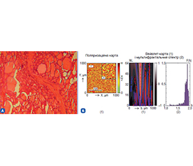
Актуальність. Розвиток захворювань щитоподібної залози (ЩЗ) є патофізіологічним взаємозв’язком між класичним і дизрегуляційним патологічними процесами, що можуть змінюватися у просторі і часі, що необхідно для своєчасної корекції технологій лікування. Мета: провести патофізіологічний аналіз дизрегуляційного і класичного патофізіологічного процесів у розвитку захворювань щитоподібної залози з можливістю оцінки ролі поляризаційної біомедичної оптики в проведенні їх діагностики. Матеріали та методи. Досліджувалися 4 групи хворих: контрольна 1-ша група — здорові донори (n = 51), дослідна 2-га група — пацієнти з вузловим зобом (n = 51), дослідна 3-тя група — пацієнти з автоімунним тиреоїдитом (АІТ) (n = 51), дослідна 4-та група — хворі на папілярний рак ЩЗ (n = 51), яким з діагностичною метою проводили пункційну біопсію ЩЗ. Паралельно з гістологічними дослідженнями із забарвленням гематоксилін-еозином використали такі методи поляризаційної біомедичної оптики: поляризаційні, вейвлет-карти і мультифрактальний спектр. Результати. Для нормальної ЩЗ карта поляризації азимутів демонструє рівномірне розподілення орієнтації волокон у гістологічному зрізі. Скелетон амплітуди вейвлет-коефіцієнтів вказує на компактну, однорідну структуру. Мультифрактальний спектр має чітко виражений максимум — це типовий показник добре організованої тканини без патологічних змін. Для вузлового зоба карта азимутів показує менш впорядковану поляризацію, з більшою хаотичністю волокон. Скелетон амплітуди вейвлет-коефіцієнтів має фрагментовану структуру, що може вказувати на деструктивні зміни або атипове утворення вузлів. Мультифрактальний спектр ширший і менш симетричний, що свідчить про підвищену складність мікроструктури, притаманну для патологічних станів. За АІТ карта азимутів поляризації демонструє помітну дезорганізацію структури, однак із ще присутніми локальними упорядкованими ділянками. Це узгоджується із хронічним запальним процесом. Скелетон амплітуди вейвлет-коефіцієнтів помірно фрагментований, що може свідчити про початкову фіброзну трансформацію та елементи гіперплазії. Мультифрактальний спектр є асиметричним, але менш вираженим у крайніх значеннях, що є натяком на фонову автоімунну активність без злоякісного росту. За папілярного раку ЩЗ карта азимутів показує суттєво хаотичну орієнтацію волокон без помітних зон впорядкування як типову ознаку злоякісної трансформації. Скелетон амплітуди вейвлет-коефіцієнтів демонструє виражену фрагментацію з глибокими порушеннями тканинної архітектоніки. Широкий мультифрактальний спектр з кількома локальними максимумами відображає високий рівень мікроструктурної складності, що характерно для онкопроцесів. Висновки. За вузлового зоба, автоімунного тиреоїдиту, папілярного раку щитоподібної залози встановлений тісний інтегративний взаємозв’язок між класичним і дизрегуляційним патологічними процесами, що наочно продемонстровано при паралельних гістологічних дослідженнях з використанням високочутливих методів поляризаційної біомедичної оптики — поляризаційної, вейвлет-карт і мультифрактального спектра.
Background. The development of thyroid diseases is a pathophysiological relationship between classical and dysregulated pathological processes that can dynamically change in space and time, which is an important need for timely correction of treatment technologies. The purpose of the study was to conduct an analysis of dysregulated and classical pathophysiological processes in the development of thyroid diseases with the possibility of assessing the role of polarization biomedical optics in its diagnosis. Materials and methods. Four groups were studied: control group 1 — healthy donors (n = 51), study group 2 — patients with nodular goiter (n = 51), study group 3 — participants with autoimmune thyroiditis (n = 51), study group 4 — patients with papillary thyroid cancer (n = 51) who underwent thyroid biopsy for diagnostic purposes. In parallel with histological studies and hematoxylin-eosin staining, the following methods of polarization biomedical optics were used: polarization, wavelet maps and multifractal spectrum. Results. For a normal thyroid gland, the azimuth polarization map shows a uniform distribution of fiber orientation in the histologic section. The amplitude skeleton of the wavelet coefficients indicates a compact, homogeneous structure. The multifractal spectrum has a distinct maximum, which is a typical indicator of well-organized tissue without pathological changes. For nodular goiter, the azimuth map shows less ordered polarization, with more chaotic fibers. The skeleton of wavelet coefficient amplitude has a fragmented structure, which may indicate destructive changes or atypical nodule formation. The multifractal spectrum is wider and less symmetrical, indicating an increased complexity of the microstructure typical for pathological conditions. In autoimmune thyroiditis, the polarization azimuth map shows a marked disorganization of the structure, but with local ordered areas still present. This is consistent with a chronic inflammatory process. The skeleton of the wavelet coefficient amplitude is moderately fragmented, which may indicate initial fibrous transformation and elements of hyperplasia. The multifractal spectrum is asymmetric, but less pronounced at the extremes, which indicates background autoimmune activity without malignant growth. In papillary thyroid cancer, the azimuth map shows a significantly chaotic orientation of fibers without noticeable zones of order — a typical sign of malignant transformation. The skeleton of the wavelet coefficient amplitude demonstrates pronounced fragmentation with deep disorders of tissue architectonics. A broad multifractal spectrum with several local maxima reflects a high level of microstructural complexity, which is characteristic of cancer. Conclusions. In case of nodular goiter, autoimmune thyroiditis, papillary thyroid cancer, a close integrative relationship between classical and dysregulated pathological processes was found, which was clearly demonstrated in parallel histological studies using highly sensitive methods of polarization biomedical optics: polarization, wavelet maps and multifractal spectrum.
вузловий зоб; автоімунний тиреоїдит; папілярний рак; щитоподібна залоза; класичний і дизрегуляційний патологічний процес; поляризаційна біомедична оптика; інтегративний підхід
nodular goiter; autoimmune thyroiditis; papillary thyroid cancer; thyroid gland; classical and dysregulated pathological process; polarization biomedical optics; integrated approach
Для ознакомления с полным содержанием статьи необходимо оформить подписку на журнал.
- Friedman Y. Conceptual scaffolding for the philosophy of me–dicine. Med Health Care Philos. 2025 Mar;28(1):45-64. doi: 10.1007/s11019-024-10231-w.
- Al-Saghbini MS, Fayyad MA, Gharaibeh L. Pathology as a Basic Medical Subject: Its Relevance and Application During Clinical Practice in Jordanian MD Programs, Interns’ and Residents’ Perspectives. Adv Med Educ Pract. 2024 Jul 4;15:627-635. doi: 10.2147/AMEP.S446158. PMID: 38983272; PMCID: PMC11230855.
- Ushenko VA, Sdobnov AYu, Mishalov WD, Dubolazov AV, Olar OV, Bachinskyi VT, et al. Biomedical applications of Jones-matrix tomography to polycrystalline films of biological fluids. Journal of Innovative Optical Health Sciences. 2019;12(06):1950017. https://doi.org/10.1142/S1793545819500172.
- Rohovyi Y, Bilookyi O, Ushenko O, Bilookyi V. The principle of direct and negative feedback regulation of endocrine functions and the possibility of using polarization biomedical optic methods in the diagnosis of nodular goiter. International Journal of Endocrinology (Ukraine). 2024;20(4):316-322. https://doi.org/10.22141/2224-0721.20.4.2024.1411.
- Rohovyi Y, Savka V, Bilookyi V, Sheremet M, Bocharov A, et al. Integrative pathophysiological and correlation-optical study of the kidneys for the formation of tubulo-interstitial syndrome: part 1 — polarization and birefringence structure. Sixteenth International Conference on Correlation Optics. 2024;12938. https://doi.org/10.1117/12.3014077.
- Ushenko A, Dubolazov A, Zheng J, Litvinenko A, Gorsky M, Ushenko Y, et al. 3D polarization-interference holographic histo–logy for wavelet-based differentiation of the polycrystalline component of biological tissues with different necrotic states. Forensic applications. J Biomed Opt. 2024 May;29(5):052920. doi: 10.1117/1.JBO.29.5.052920. Epub 2024 Mar 15. PMID: 38495527; PMCID: PMC10943250.
- Zhengbing Hu, Tereikovskyi I, Chernyshev D, Tereikovska L, Tereikovskyi O, Dong Wang. Procedure for Processing Biometric Parameters Based on Wavelet Transformations. International Journal of Modern Education and Computer Science (IJMECS). 2021;13(2):11-22. doi: 10.5815/ijmecs.2021.02.02.
- Rohovyi Y, Bilookyi O, Ushenko O, Bilookyi V, Semenenko S. The role of histohematologic barriers and the possibility of using polarization biomedical optics methods in the diagnosis of autoimmune thyroiditis. International Journal of Endocrinology (Ukraine). 2024;20(6):452-458. https://doi.org/10.22141/2224-0721.20.6.2024.1442.
- Wong KS, Angell TE, Barletta JA, Krane JF. Hürthle cell lesions of the thyroid: Progress made and challenges remaining. Cancer Cytopathol. 2021 May;129(5):347-362. doi: 10.1002/cncy.22375. Epub 2020 Oct 27. PMID: 33108684.
- McFadden DG, Sadow PM. Genetics, Diagnosis, and Mana–gement of Hürthle Cell Thyroid Neoplasms. Front Endocrinol (Lausanne). 2021 Jun 10;12:696386. doi: 10.3389/fendo.2021.696386. PMID: 34177816; PMCID: PMC8223676.
- Brix K, Szumska J, Weber J, Qatato M, Venugopalan V, Al-Hashimi A, et al. Auto-Regulation of the Thyroid Gland Beyond Classical Pathways. Exp Clin Endocrinol Diabetes. 2020 Jun;128(6-07):437-445. doi: 10.1055/a-1080-2969. Epub 2020 Feb 19. PMID: 32074633.
- Kenarlı K, Bahçecioğlu AB, Aksu ÖB, Güllü S. Are sonographic characteristics of Hashimoto’s thyroiditis related with immunologic parameters? A cross-sectional study. J Endocrinol Invest. 2024 Jul;47(7):1701-1709. doi: 10.1007/s40618-023-02286-y. Epub 2024 Jan 21. PMID: 38245884.
- Napolitano G, Bucci I, Di Dalmazi G, Giuliani C. Non-Conventional Clinical Uses of TSH Receptor Antibodies: The Case of Chronic Autoimmune Thyroiditis. Front Endocrinol (Lausanne). 2021 Nov 5;12:769084. doi: 10.3389/fendo.2021.769084. PMID: 34803929; PMCID: PMC8602826.
- Nguyen CT, Singer PA, Nguyen T. Thyroiditis — a clinical update and review. TTU J Biomed Sci. 2023;2:33-40. https://doi.org/10.53901/tjbs.2023.08.art04.
- Vargas-Uricoechea H, Castellanos-Pinedo A, Urrego-Nogue–ra K, Pinzón-Fernández MV, Meza-Cabrera IA, Vargas-Sierra H. A Scoping Review on the Prevalence of Hashimoto’s Thyroiditis and the Possible Associated Factors. Med Sci (Basel). 2025 Apr 10;13(2):43. doi: 10.3390/medsci13020043. PMID: 40265390; PMCID: PMC12015930.
- Wang C, Ma W, Qin L, Wu L, Liu T. Efficacy of Xiaoyao-san preparations in treating Hashimoto’s thyroiditis: a meta-analysis and systematic review. Front Pharmacol. 2025 Jun 13;16:1528506. doi: 10.3389/fphar.2025.1528506. PMID: 40584613; PMCID: PMC12202410.
- Abdalrahman S, Smail HO, Shallal AF. Genetic and epigene–tic markers in Hashimoto’s thyroiditis: a comprehensive review. Int J Epigenet. 2025 Jan;5(1):1-20. doi: 10.3892/ije.2025.24.
- Zhang Q, Lan X. Assessment of causal association between autoimmune thyroiditis and thyroid cancer: A Mendelian rando–mization study. Medicine (Baltimore). 2025 Feb 28;104(9):e41633. doi: 10.1097/MD.0000000000041633. PMID: 40020149; PMCID: PMC11875592.
- Zhao Z, Gao Y, Pei X, Wang W, Zhang H. Causal role of immune cells in Hashimoto’s thyroiditis: Mendelian randomization study. Front Endocrinol (Lausanne). 2024 May 13;15:1352616. doi: 10.3389/fendo.2024.1352616. PMID: 38803479; PMCID: PMC11128540.
- Gubbiotti MA, Canberk S, Baloch ZW. Oncocytic Tumors in the Thyroid: A Tri-Focal Review — Integrated Cytopathological, Pathological, and Molecular Perspectives. Acta Cytol. 2025 Feb 14:1-17. doi: 10.1159/000544739. Epub ahead of print. PMID: 39956101.
- Asa SL, Mete O. Oncocytic Change in Thyroid Pathology. Front Endocrinol (Lausanne). 2021 May 3;12:678119. doi: 10.3389/fendo.2021.678119. PMID: 34012422; PMCID: PMC8127945.
- Lebrun L, Salmon I. Pathology and new insights in thyroid neoplasms in the 2022 WHO classification. Curr Opin Oncol. 2024 Jan 1;36(1):13-21. doi: 10.1097/CCO.0000000000001012. Epub 2023 Nov 17. PMID: 37975316; PMCID: PMC10715705.
- Wang X, Liu Y, Chen L, Zhang J, Jiang R, Zhang L, et al. Oncocytic cell carcinoma of the thyroid with TERT promoter mutation presenting as asphyxia in an elderly: a case report. Front Endocrinol (Lausanne). 2024 Aug 16;15:1349114. doi: 10.3389/fendo.2024.1349114. PMID: 39220363; PMCID: PMC11362092.