
Международный эндокринологический журнал Том 21, №5, 2025
Вернуться к номеру
Аналіз дизрегуляційного і класичного патофізіологічних процесів за розвитку автоімунного тиреоїдиту
Авторы: Роговий Ю.Є., Білоокий О.В., Білоокий В.В., Савчук Т.П.
Буковинський державний медичний університет, м. Чернівці, Україна
Рубрики: Эндокринология
Разделы: Клинические исследования
Версия для печати
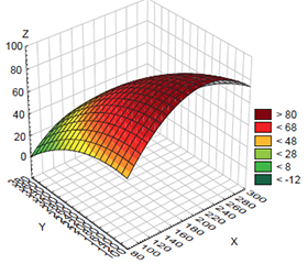
Актуальність. Розвиток гіпертрофічної, атрофічної і фіброзної форм автоімунного тиреоїдиту (АІТ) являє собою динамічний взаємозв’язок між класичним і дизрегуляційним патологічними процесами, які можуть швидко змінюватися у просторі і часі, що важливо для своєчасної корекції технології лікування. Мета: провести аналіз та оцінити роль дизрегуляційного і класичного патофізіологічних процесів у розвитку гіпертрофічної, атрофічної і фіброзної форм автоімунного тиреоїдиту. Матеріали та методи. Досліджувалися три групи хворих: група 1 — хворі на гіпертрофічний АІТ (n = 21), група 2 — пацієнти з атрофічним АІТ (n = 18), група 3 — хворі на фіброзний АІТ (n = 12), яким з діагностичною метою проводили тонкоголкову аспіраційну пункційну біопсію щитоподібної залози (ЩЗ). Отриманий матеріал досліджували гістологічно шляхом забарвлення гематоксилін-еозином. За допомогою імуноферментного аналізу визначали тиреотропний гормон (ТТГ), вільний трийодтиронін (вТ3), вільний тироксин (вТ4), тиреоглобулін (ТГ), антитіла до тиреоглобуліну (АТ-ТГ), антитіла до тиреоїдної пероксидази (АТ-ТПО). При статистичному аналізі результатів дослідження вираховувалися середня арифметична вибірки (x), стандартна помилка середньої арифметичної (Sx), вірогідність відмінностей між групами оцінювали за допомогою непараметричного критерію Манна — Уїтні (р < 0,05). Використовувався багатофакторний регресійний аналіз за допомогою програми Statistica. Результати. Встановлено вірогідно вищий рівень ТТГ при атрофічному і фіброзному АІТ порівняно з гіпертрофічною формою АІТ. Рівень вТ4 при зазначених формах АІТ був нижчим щодо гіпертрофічного АІТ. Водночас рівень вТ3 був зниженим тільки при фіброзному АІТ порівняно з атрофічним. АТ-ТПО були нижчими при атрофічному і фіброзному АІТ порівняно з гіпертрофічним АІТ. АТ-ТГ були нижчими тільки при атрофічному АІТ. Проведення кореляційного аналізу виявило вірогідні кореляційні зв’язки: позитивний — в парі ТТГ — вТ4 та негативні — в парах ТТГ — ТГ, Т4 — ТГ, АТ-ТГ — ТГ за гіпертрофічного АІТ. Кореляційний аналіз за атрофічного АІТ показав вірогідні кореляційні зв’язки: позитивні — в парах АТ-ТПО — ТГ, АТ-ТГ — ТГ та негативні — в парах ТТГ — ТГ, ТТГ — АТ-ТПО. Кореляційний аналіз встановив вірогідні кореляційні зв’язки: позитивний — в парі АТ-ТПО — АТ-ТГ та негативні — в парах вТ4 — ТГ, вТ3 — АТ-ТПО за фіброзного АІТ. Висновки. За умов розвитку автоімунного тиреоїдиту встановлений динамічний взаємозв’язок між класичним і дизрегуляційним патологічними процесами, який містить три стадії: гіпертрофічний, атрофічний і фіброзний автоімунний тиреоїдит, що призводить до формування великого порочного кола патогенезу внаслідок дизрегуляційної активації клітин Гюртле — Ашкеназі.
Background. The development of hypertrophic, atrophic and fibrous autoimmune thyroiditis (AIT) is a dynamic relationship between classical and dysregulatory pathological processes that can change rapidly in space and time, which is important for timely correction of treatment technology. Aim of the study was to analyze and evaluate the role of dysregulatory and classical pathophysiological processes in the development of hypertrophic, atrophic and fibrous autoimmune thyroiditis. Materials and methods. Three groups of patients were studied: group 1 — with hypertrophic AIT (n = 21), group 2 — atrophic AIT (n = 18), group 3 — fibrous AIT (n = 12), who underwent a fine needle aspiration biopsy of the thyroid gland for diagnostic purposes. The obtained material was examined histologically by hematoxylin-eosin staining. Enzyme immunoassay was used to determine thyroid-stimulating hormone (TSH), free triiodothyronine (fT3), free thyroxine (fT4), thyroglobulin (TG), antibodies to thyroglobulin (TG-Ab), and antibodies to thyroid peroxidase (TPO-Ab). In the statistical analysis of the study results, the arithmetic mean of the sample, the standard error of the arithmetic mean were calculated, and the significance of differences between the groups was assessed using the nonparametric Mann-Whitney test (p < 0.05). Multivariate regression analysis was performed using the Statistica program. Results. TSH levels were found to be significantly higher in atrophic and fibrous AIT compared to hypertrophic form. Content of fT4 in these nosologies was lower than in hypertrophic AIT. At the same time, fT3 was reduced only in fibrous AIT compared to atrophic type. TPO-Ab were lower in atrophic and fibrous AIT than in hypertrophic AIT. TG-Ab were lower only in atrophic AIT. The correlation analysis revealed significant correlations: positive in the TSH — fT4 pair and negative in the TSH — TG, fT4 — TG, TG-Ab — TG pairs in hypertrophic AIT. In atrophic AIT, the correlation analysis showed significant correlations: positive in the pairs TPO-Ab — TG, TG-Ab — TG and negative in the pairs TSH — TG, TSH — TPO-Ab. The correlation analysis revealed reliable correlations: positive in the pair TPO-Ab — TG-Ab and negative in the pairs fT4 — TG, fT3 — TPO-Ab in fibrous AIT. Conclusions. In case of autoimmune thyroiditis, a dynamic relationship was found between classical and dysregulatory pathological processes, which includes three stages: hypertrophic, atrophic, and fibrous autoimmune thyroiditis, leading to the formation of a large vicious circle of pathogenesis due to dysregulated activation of Hürthle-Askanazy cells.
автоімунний тиреоїдит; прямі та негативні зворотні зв’язки; щитоподібна залоза; класичний і дизрегуляційний патологічний процес; велике порочне коло, клітини Гюртле — Ашкеназі
autoimmune thyroiditis; direct and negative feedback; thyroid gland; classical and dysregulatory pathological process; large vicious circle; Hürthle-Askanazy cells
Для ознакомления с полным содержанием статьи необходимо оформить подписку на журнал.
- Reich AD, Hansen HB, Link BG. Fundamental Interventions: How Clinicians Can Address the Fundamental Causes of Disease. J Bioeth Inq. 2016 Jun;13(2):185-92. doi: 10.1007/s11673-016-9715-3. Epub 2016 Mar 29. PMID: 27022923; PMCID: PMC5540132.
- Fundamentals of the Theory of Medicine: monograph. A.I. Gozhenko, V.S. Biryukov, O.A. Gozhenko, et al.; under the general editorship of A.I. Gozhenko. Odesa: Fenix, 2024. 248 p. (in Ukrainian)
- Vinay Kumar (Editor), Abul K Abbas (Editor), Jon C Aster (Edi–tor), Andrea T Deyrup (Editor), Abhijit Das (Editor), Stanley L Robbins. Robbins Basic Pathology. 2023, 11th ed. https://www.worldcat.org/title/1357077633.
- Khlamanova LI, Tkachenko YuV, Severylova MD. Structural and Functional Characteristics of Different Histohematological Barriers of the Organism in Norm and during the Pathological Changes, their Medical Significance and Role in Forming Clinical Thinking of Junior Students. Ukrainian Journal of Medicine, Biology and Sports. 2018;1(10):245-252. DOI: 10.26693/jmbs03.01.245. (in Ukrainian)
- Ralli M, Angeletti D, Fiore M, D’Aguanno V, Lambiase A, Artico M, et al. Hashimoto’s thyroiditis: An update on pathogenic mecha–nisms, diagnostic protocols, therapeutic strategies, and potential malignant transformation. Autoimmun Rev. 2020 Oct;19(10):102649. doi: 10.1016/j.autrev.2020.102649. Epub 2020 Aug 15. PMID: 32805423.
- Kenarlı K, Bahçecioğlu AB, Aksu ÖB, Güllü S. Are sonographic characteristics of Hashimoto’s thyroiditis related with immunologic parameters? A cross-sectional study. J Endocrinol Invest. 2024 Jul;47(7):1701-1709. doi: 10.1007/s40618-023-02286-y. Epub 2024 Jan 21. PMID: 38245884.
- Rohovyi Y, Bilookyi O, Ushenko O, Bilookyi V, Semenenko S. The role of histohematologic barriers and the possibility of using polarization biomedical optics methods in the diagnosis of autoimmune thyroiditis. International Journal оf Endocrinology (Ukraine). 2024;20(6):452-458. https://doi.org/10.22141/2224-0721.20.6.2024.1442.
- Landers K, Richard K. Traversing barriers — How thyroid hormones pass placental, blood-brain and blood-cerebrospinal fluid barriers. Mol Cell Endocrinol. 2017 Dec 15;458:22-28. doi: 10.1016/j.mce.2017.01.041. Epub 2017 Jan 30. PMID: 28153799.
- Brix K, Szumska J, Weber J, Qatato M, Venugopalan V, Al-Hashimi A, et al. Auto-Regulation of the Thyroid Gland Beyond Classical Pathways. Exp Clin Endocrinol Diabetes. 2020 Jun;128(6-07):437-445. doi: 10.1055/a-1080-2969. Epub 2020 Feb 19. PMID: 32074633.
- Caroline T. Nguyen, Peter A. Singer, Thach Nguyen. Thyroiditis — A Clinical Update and Review TTU. Journal of Biomedical Sciences. 2023;02:33-40. DOI: 10.53901/ tjbs.2023.08.art04.
- Napolitano G, Bucci I, Di Dalmazi G, Giuliani C. Non-conventional clinical uses of TSH receptor antibodies in chronic autoimmune thyroiditis. Front Endocrinol. 2021;12:769084. https://doi.org/10.3389/fendo.2021.769084.
- Vargas‑Uricoechea H, Castellanos‑Pinedo A, Urrego‑Nogue–ra K, Pinzón‑Fernández MV, Meza‑Cabrera IA, Vargas‑Sierra H. A scoping review on the prevalence of Hashimoto’s thyroiditis and the possible associated factors. Med Sci (Basel). 2025 Apr 10;13(2):43. doi: 10.3390/medsci13020043.
- Chen Y, Li J, Zhang H, et al. Systematic review of supplements targeting thyroid autoimmunity in Hashimoto’s thyroiditis. Front Endocrinol (Lausanne). 2024 Dec;15:1445878. doi: 10.3389/fendo.2024.1445878.
- Abdalrahman S, Smail HO, Shallal AF. Genetic and epige–netic markers in Hashimoto’s thyroiditis: a comprehensive review. Int J Epigenet. 2025 Jan;5(1):1-20. doi: 10.3892/ije.2025.24.
- Lim J, Hu H, Kim S, et al. Causal role of immune cell traits in Hashimoto’s thyroiditis: a Mendelian randomization study. Front Endocrinol (Lausanne). 2024 Mar;15:1352616. doi: 10.3389/fendo.2024.1352616.
- Chen Z, van der Sman ASE, Groeneweg S, de Rooij LJ, Visser WE, Peeters RP, et al. Thyroid Hormone Transporters in a Human Placental Cell Model. Thyroid. 2022 Sep;32(9):1129-1137. doi: 10.1089/thy.2021.0503. Epub 2022 Jul 11. PMID: 35699060; PMCID: PMC9526468.
- Asa SL, Mete O. Oncocytic change in thyroid pathology: update and diagnostic implications. Front Endocrinol (Lausanne). 2021 May;12:678119. doi: 10.3389/fendo.2021.678119.
- Cannon J. The significance of Hurthle cells in thyroid di–sease. Oncologist. 2011;16(10):1380-7. doi: 10.1634/theoncolo–gist.2010-0253. Epub 2011 Sep 30. PMID: 21964000; PMCID: PMC3228061.
- Wong KS, Angell TE, Barletta JA, Krane JF. Hürthle cell lesions of the thyroid: progress made and challenges remaining. Cancer Cytopathol. 2021;129(5):347-362. https://doi.org/10.1002/cncy.22375.
- McFadden DG, Sadow PM. Genetics, diagnosis, and management of Hürthle cell thyroid neoplasms. Front Endocrinol (Lausanne). 2021;12:696386. doi: 10.3389/fendo.2021.696386.
- Gubbiotti MA, Canberk S, Baloch ZW. Oncocytic tumours in the thyroid: a tri‑focal review — integrated cytopathological, patholo–gical, and molecular perspectives. Acta Cytol. 2025 Feb 14:1-17. doi: 10.1159/000544739.
- Ghossein RA, Sobrinho-Simões M, Tallini G, et al. Oncocytic carcinoma of the thyroid gland: WHO classification and mole–cular insights. J Clin Endocrinol Metab. 2025 May;110(5):e1343. doi: 10.1210/jc.2025-e1343.
- Falhammar H, Juhlin CC, Barner C, Catrina SB, Karefylakis C, Calissendorff J. Riedel’s thyroiditis: clinical presentation, treatment and outcomes. Endocrine. 2018 Apr;60(1):185-192. doi: 10.1007/s12020-018-1526-3. Epub 2018 Jan 29. PMID: 29380231; PMCID: PMC5845586.