
Журнал «Медицина неотложных состояний» Том 21, №4, 2025
Вернуться к номеру
Оцінка міофібробластної активності на етапах хірургічного лікування ранового дефекту при ускладнених формах хронічної венозної недостатності
Авторы: Саволюк С.І. (1), Дембіцький А.Р. (1), Шмигіна І.М. (2)
(1) - Національний університет охорони здоров’я України імені П.Л. Шупика, м. Київ, Україна
(2) - Інститут післядипломної освіти Національного медичного університету ім. О.О. Богомольця, м. Київ, Україна
Рубрики: Медицина неотложных состояний
Разделы: Клинические исследования
Версия для печати
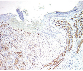
Мета роботи: оцінити загоювальний потенціал трофічних виразок (ТВ) у пацієнтів із хронічною венозною недостатністю (ХВН) С6 шляхом аналізу експресії маркера -SMA як прогностичного індикатора ефективності лікування з використанням плазми, збагаченої тромбоцитами (PRP), порівняно з традиційними методами лікування ТВ. Матеріали та методи. У дослідженні, проведеному в 2021–2023 рр., брали участь 33 пацієнти, розділені на основну (n = 15) і контрольну (n = 18) групи. Основній групі проводили термічну абляцію стовбурів підшкірних вен і місцеве лікування ТВ за допомогою PRP. У контрольній групі виконувався стрипінг неспроможних стовбурів із застосуванням традиційних засобів для місцевого лікування ТВ (мазі, пов’язки). Ефективність лікування оцінювали за клінічними параметрами, терміном загоєння ТВ та експресією -SMA у зразках тканин. Результати. Застосування PRP сприяло прискоренню епітелізації ТВ, зниженню запальних маркерів і підвищенню експресії -SMA. Це вказує на активізацію міофібробластів, що асоціюється з прискоренням загоєння ран. Порівняно з традиційними методами лікування застосування PRP у ділянці ТВ виявилося більш ефективним та привело до значного скорочення термінів загоєння ТВ. Застосування PRP у поєднанні із сучасними рановими покриттями дозволило досягти загоєння ТВ на фоні ХВН С6 на 32,9 дня (p-value = 1,044e-10), у пацієнтів групи порівняння загоєння ТВ спостерігалося на 52,7 дня (p-value = 1,044e-10). Висновки. Результати дослідження підтверджують ефективність ін’єкцій PRP у поєднанні із сучасними рановими покриттями для прискорення загоєння трофічних виразок у пацієнтів із ХВН C6.
Background. The purpose of the study was to evaluate the healing potential of trophic ulcers (TUs) in patients with chronic venous insufficiency (CVI) C6 by analyzing the expression of the -SMA marker as a prognostic indicator of treatment effectiveness using platelet-rich plasma (PRP), in comparison with traditional TU treatment methods. Materials and methods. The study conduc-ted in 2021–2023 involved 33 patients divided into the main (n = 15) and the control groups (n = 18). The main group underwent thermal ablation of the saphenous vein trunks and local treatment of TUs with PRP. In the control group, stripping of the incompetent trunks was performed using traditional local treatment for TUs (ointments, dressings). Treatment effectiveness was evaluated based on clinical parameters, TU healing time, and -SMA expression in tissue samples. Results. The use of PRP promoted faster epithelialization of TUs, reduced inflammatory markers, and increased -SMA expression. This indicates myofibroblast activation, which is associated with accelerated wound healing. Compared to traditional treatment methods, the use of PRP in the TU area proved to be more effective and resulted in a significant reduction in TU healing time. The application of PRP combined with modern wound dressings allowed to achieve TU healing in CVI C6 patients by day 32.9 (p-value = 1.044e-10), whereas in the comparison group, TU hea-ling was observed by day 52.7 (p-value = 1.044e-10). Conclusions. The results of the study confirm the effectiveness of PRP injections in combination with modern wound dressings to accelerate the healing of trophic ulcers in patients with CVI C6.
хронічна венозна недостатність; трофічна виразка; імуногістохімія; -SMA; плазма, збагачена тромбоцитами
chronic venous insufficiency; trophic ulcer; immunohistochemistry; -SMA; platelet-rich plasma
Для ознакомления с полным содержанием статьи необходимо оформить подписку на журнал.
- Kestler, B. (2021). Chronic Venous Insufficiency. Physician Assistant Clinics, 6, 319-330. https://doi.org/10.1016/j.cpha.2020.11.005.
- Mansilha, A. (2020). Early Stages of Chronic Venous Disease: Medical Treatment Alone or in Addition to Endovenous Treatments. Advances in Therapy, 37, 13-18. https://doi.org/10.6084/m9.figshare.11417631.
- Nicolaides, A., Kakkos, S., Baekgaard, N., Comerota, A., De Maeseneer, M., Eklof, B., et al. (2018). Management of chronic Venous disorders of the Lower Limbs. Guidelines According to Scientific Evidence, part I, 181-254.
- Brand, F., Dannenberg, A., Abbott, R. and Kannel, W. (1988). The Epidemiology of Varicose Veins: The Framingham Study. Am J Prev Med, 4, 96-101. https://pubmed.ncbi.nlm.nih.gov/3395496/.
- Pannier, F. and Rabe, E. (2012). The Relevance of the Natural History of Varicose Veins and Refunded Care. Phlebology, 27, 23-26. https://doi.org/10.1258/PHLEB.2012.012S23.
- Berenguer Prez, M., Lpez-Casanova, P., Sarabia Lavn, R., Gonzlez de la Torre, H. and Verd-Soriano, J. (2019). Epidemiology of Venous Leg Ulcers in Primary Health Care: Incidence and Prevalence in a Health Centre — A Time Series Study (2010-2014). Int Wound J, 16, 256-265. https://doi.org/10.1111/IWJ.13026.
- Gonzlez-Consuegra, R.V. and Verd, J. (2011). Quality of Life in People with Venous Leg Ulcers: An Integrative Review. J Adv Nurs, 67, 926-944. https://doi.org/10.1111/J.1365-2648.2010.05568.X.
- Probst, S., Schaud, L., Bobbink, P., Skinner, M.B. and Weller, C.D. (2020). The Lived Experience of Recurrence Prevention in Patients with Venous Leg Ulcers: An Interpretative Phenomenological Study. J Tissue Viability, 29, 176-179. https://doi.org/10.1016/J.JTV.2020.01.001.
- Krber, A., Klode, J., Al-Benna, S., Wax, C., Schadendorf, D., Steinstraesser, L. and Dissemond, J. (2011). Etiology of Chronic Leg Ulcers in 31,619 Patients in Germany Analyzed by an Expert Survey. J Dtsch Dermatol Ges, 9, 116-121. https://doi.org/10.1111/J.1610-0387.2010.07535.X.
- Krber, A., Jockenhfer, F., Sondermann, W., Stoffels-Weindorf, M. and Dissemond, J. (2017). [First Manifestation of Leg Ulcers : Analysis of Data from 1000 Patients]. Der Hautarzt; Zeitschrift fur Dermatologie, Venerologie, und verwandte Gebiete, Hautarzt, 68, 483-491. https://doi.org/10.1007/S00105-017-3950-3.
- Rajhathy, E.M., Murray, H.D., Roberge, V.A. and Woo, K.Y. (2020). Healing Rates of Venous Leg Ulcers Managed With Compression Therapy: A Secondary Analysis of Data. J Wound Ostomy Continence Nurs, 47, 477-483. https://doi.org/10.1097/WON.0000000000000693.
- Probst, S., Saini, C., Gschwind, G., Stefanelli, A., Bobbink, P., Pugliese, M.T., et al. (2023). Prevalence and Incidence of Venous Leg Ulcers — A Systematic Review and Meta-Analysis. International Wound Journal, 20, 3906-3921. https://doi.org/10.1111/IWJ.14272.
- Fernandes Abbade, L.P. and Lastria, S. (2005). Venous Ulcer: Epidemiology, Physiopathology, Diagnosis and Treatment. Int J Dermatol, 44, 449-456. https://doi.org/10.1111/J.1365-4632.2004.02456.X.
- Nussbaum, S.R., Carter, M.J., Fife, C.E., DaVanzo, J., Haught, R., Nusgart, M. and Cartwright, D. (2018). An Economic Evaluation of the Impact, Cost, and Medicare Policy Implications of Chronic Nonhealing Wounds. Value Health, 21, 27-32. https://doi.org/10.1016/J.JVAL.2017.07.007.
- Urwin, S., Dumville, J.C., Sutton, M. and Cullum, N. (2022). Health Service Costs of Treating Venous Leg Ulcers in the UK: Evidence from a Cross-Sectional Survey Based in the North West of England. BMJ Open, 12. https://doi.org/10.1136/BMJOPEN-2021-056790.
- Weller, C. and Evans, S. (2012). Venous Leg Ulcer Management in General Practice — Practice Nurses and Evidence Based Guidelines. Aust Fam Physician, 41, 331-337. https://pubmed.ncbi.nlm.nih.gov/22558626/.
- Kamhawy, A.H., Elbarbary, A.H., Elhenidy, M.A. and Elwagih, A.M.M. (2020). Periulcer Foam Sclerotherapy Injection in Chronic Venous Leg Ulcers Using Near-Infrared Laser for Vein Visualization. Int J Low Extrem Wounds, 19, 63-69. https://doi.org/10.1177/1534734619870680.
- Piccin, A., Di Pierro, A.M., Canzian, L., Primerano, M., Corvetta, D., Negri, G., et al. (2017). Platelet Gel: A New Therapeutic Tool with Great Potential. Blood Transfus, 15, 333-340. https://doi.org/10.2450/2016.0038-16.
- Elsawy, A. and Abdelraouf, H. (2021). Effect of Ultrasound-Guided Injection of Local Ozone or Platelet Rich Plasma Versus Corticosteroid in Plantar Fasciitis. Al-Azhar International Medical Journal, 1, 224-230. https://doi.org/10.21608/AIMJ.2021.52095.1368.
- Alshahat, O., Taha, A. and Abdulaziz, M. (2020). Assessment the Role of Platelet Rich Plasma in Follicular Unit Extraction Hair Transplantation. Al-Azhar International Medical Journal, 1, 1-12. https://doi.org/10.21608/AIMJ.2020.29351.1217.
- Еl tawab, W.A., Hammoda, I. and Zayed, E. (2020). The Effectiveness of Platelet-Rich Plasma Injection in Management of Rotator Cuff Tendinopathy. Al-Azhar International Medical Journal, 1, 193-196. https://doi.org/10.21608/aimj.2021.49733.1351.
- Werner, S., Krieg, T. and Smola, H. (2007). Keratinocyte-Fibroblast Interactions in Wound Healing. J Invest Dermatol, 127, 998-1008. https://doi.org/10.1038/SJ.JID.5700786.
- Darby, I.A., Laverdet, B., Bont, F. and Desmoulire A. (2014). Fibroblasts and Myofibroblasts in Wound Healing. Clinical, Cosmetic and Investigational Dermatology, 7, 301-311. https://sci-hub.se/10.2147/ccid.s50046.
- Roosterman, D., Goerge, T., Schneider, S.W., Bunnett, N.W. and Steinhoff, M. (2006). Neuronal Control of Skin Function: The Skin as a Neuroimmunoendocrine Organ. Physiol Rev, 86, 1309-1379. https://doi.org/10.1152/PHYSREV.00026.2005.
- Hu, Z., Wang, S., Yang, H., Xv, H., Shan, B., Lin, L. and Han, X. (2024). Efficacy and Safety of Platelet-Rich Plasma in the Treatment of Venous Ulcers: A Systematic Review and Meta-Analysis of Randomized Controlled Trials. International Wound Journal, 21, 1-15. https://doi.org/10.1111/IWJ.14736.
- Elbarbary, A.H., Hassan, H.A. and Elbendak, E.A. (2020). Autologous Platelet-rich Plasma Injection Enhances Healing of Chronic Venous Leg Ulcer: A Prospective Randomised Study. International Wound Journal, 17, 992. https://doi.org/10.1111/IWJ.13361.
- Kushida, S., Kakudo, N., Suzuki, K. and Kusumoto, K. (2013). Effects of Platelet-Rich Plasma on Proliferation and Myofibroblastic Differentiation in Human Dermal Fibroblasts. Ann Plast Surg, 71, 219-224. https://doi.org/10.1097/SAP.0B013E31823CD7A4.
- Chellini, F., Tani, A., Vallone, L., Nosi, D., Pavan, P., Bambi, F., et al. (2018). Platelet-Rich Plasma Prevents in Vitro
- Transforming Growth Factor-1-Induced Fibroblast to Myofibroblast Transition: Involvement of Vascular Endothelial Growth Factor (VEGF)-A/VEGF Receptor-1-Mediated Signaling. Cells, 7, 142. https://doi.org/10.3390/CELLS7090142.
- Yilmaz, S., Aksoy, E., Doganci, S., Yalcinkaya, A., Di-ken, A.I. and Cagli, K. (2015). Autologous Platelet-Rich Plasma in Treatment of Chronic Venous Leg Ulcers: A Prospective Case Series. Vascular, 23, 580-585. https://doi.org/10.1177/1708538114563824.