Международный эндокринологический журнал Том 21, №1, 2025
Вернуться к номеру
Динаміка гормональних та імунологічних показників у хворих на гостру емпієму плеври
Авторы: V.V. Tkachenko (1, 3), V.V. Boyko (2, 3), V.V. Kritsak (1, 3), A.L. Sochnieva (1), D.V. Minukhin (2), A.O. Merkulov (3), V.G. Hroma (2), V.V. Ponomarova (1), O.P. Sharmazanova (1)
(1) - Medical Institute, National Technical University “Kharkiv Polytechnic Institute”, Kharkiv, Ukraine
(2) - Kharkiv National Medical University, Kharkiv, Ukraine
(3) - V.T. Zaytsev Institute of General and Urgent Surgery of NAMSU, Kharkiv, Ukraine
Рубрики: Эндокринология
Разделы: Клинические исследования
Версия для печати
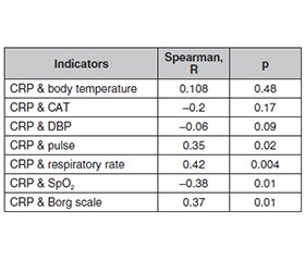
Актуальність. При гострій емпіємі плеври виявляють виражені порушення клітинних і гуморальних факторів імунітету, а також неспецифічної резистентності організму. Мета: аналіз і визначення результатів гормональних та імунологічних досліджень в осіб із гострою емпіємою плеври залежно від тяжкості перебігу захворювання. Матеріали та методи. Вивчено динаміку гормональних та імунологічних змін у 64 хворих на гостру емпієму плеври, яких лікували за допомогою класичних і малоінвазивних хірургічних методів. Учасники були розподілені на 5 груп за ступенем тяжкості захворювання, що визначали за критеріями, які включали наступні клініко-лабораторні показники: частота дихання, пульсу, артеріальний тиск, температурна реакція, кількість уражених часток, лейкоцитоз, SpO2. В усіх пацієнтів проводили стандартне обстеження, а також вивчали сироваткову концентрацію інтерлейкіну (IЛ) 6, IЛ-8, фактора некрозу пухлини α (ФНП-α), С-реактивного білка (СРБ). Результати. Оцінка цитокінового спектра у хворих на гостру емпієму плеври дозволяє констатувати стан гіперцитокінемії з підвищенням прозапальних цитокінів. При цьому ступінь останнього відрізнявся залежно від тяжкості хвороби. Вивчення структури цитокінового статусу дозволило встановити, що у хворих із тяжким перебігом емпієми плеври спостерігалося вірогідне зростання концентрації ІЛ-6 та СРБ. Проте тяжкий перебіг захворювання пов’язаний із недостатнім підвищенням IЛ-8 і ФНП-α. У пацієнтів старшої вікової групи виражений дефіцит IЛ-8 і меншою мірою IЛ-6. У загальному аналізі крові хворих із тяжким перебігом захворювання виявлено збільшення загальної кількості лейкоцитів, нейтрофілів, виражений зсув лейкоцитарної формули вліво, підвищення швидкості осідання еритроцитів. Висновки. Визначено фактори, що впливають на тяжкий перебіг гострої емпієми плеври: зниження насичення киснем менше 94 %, вираженість задишки, що перевищує 2 бали за шкалою Борга, ураження 3 і більше сегментів легеневої тканини, а також численні клінічні ознаки порушення протиінфекційного захисту, виражене зниження рівня нейтрофілів, підвищення концентрації СРБ та недостатнє підвищення IЛ-8 і ФНП-α в сироватці крові.
Background. In case of acute pleural empyema, pronounced violations of cellular and humoral factors of immunity, as well as non-specific resistance of the body are revealed. The purpose of our research: to analyze and identify results of hormonal and immunological research in patients with acute pleural empyema depending on the severity of disease. Materials and methods. Dynamics of hormonal and immunological changes has been studied in 64 participants with acute pleural empyema who were treated with classical and minimally invasive surgical methods. Patients were classified into 5 groups depending on disease severity determined according to the criteria which included clinical and laboratory parameters, such as respiratory rate, heart rate, blood pressure, temperature reaction, the number of affected particles, leukocytosis, SpO2. All the patients underwent a standard examination, as well as determination of serum concentration of interleukin (IL) 6, IL-8, tumor necrosis factor (TNF) α, C-reactive protein (CRP). Results. Assessment of blood cytokines in patients with acute pleural empyema allows detecting hypercytokinemia with an increase in pro-inflammatory cytokines. Meanwhile, the degree of their elevation differed depending on the disease severity. Study of structure of cytokine status allowed us to identify that patients with severe course of pleural empyema had a significant increase in IL-6 and CRP. Nevertheless, the severe course of the disease is associated with insufficient increase of IL-8 and TNF-α. Patients of the older age group have deficiency of IL-8 and to a lesser extent IL-6. Complete blood count revealed higher total number of leukocytes, neutrophils, pronounced left shift, increased erythrocyte sedimentation rate in patients with a severe course of the disease. Conclusions. There have been identified factors which affect severe course of acute pleural empyema: a decrease in oxygen saturation to less than 94 %, severity of dyspnea which exceeds 2 points on the Borg scale, damage to 3 or more segments of lung tissue, and also numerous clinical signs of violation of anti-infective protection, pronounced decrease in neutrophils, an increase in the concentration of CRP and insufficient increase of IL-8 and TNF-α in blood serum.
гостра емпієма плеври; гормональні та імунологічні порушення
acute pleural empyema; hormonal and immunological disorders
Для ознакомления с полным содержанием статьи необходимо оформить подписку на журнал.
- Finocchiaro G, Kobayashi Y, Magavern E, et al. Prevalence and prognostic role of right ventricular involvement in stress-induced cardiomyopathy. J Card Fail. 2015;21(5):419-425. doi: 10.1016/j.cardfail.2015.02.001.
- Ambrogi MC, Fanucchi O, Melfi F, Mussi A. Robotic surgery for lung cancer. The Korean journal of thoracic and cardiovascular surgery. 2014;47(3):201-210. doi: 10.5090/kjtcs.2014.47.3.201.
- Barthwal MS, Marwah V, Chopra M, et al. A Five-Year Study of Intrapleural Fibrinolytic Therapy in Loculated Pleural Collections. Indian J Chest Dis Allied Sci. 2016;58(1):17-20.
- Boyko VV, Lopatenko DE. Pathogenic flora in pyopneumothorax and its sensitivity to antibiotics. Kharkiv surgical school. 2013;4(61):54-56.
- Eliashar R, Davros W, Eliachar I. Virtual endoscopy of the upper airway — a diagnostic tool. Postgrad Med J. 2000;76(893):187-188. doi: 10.1136/pmj.76.893.187.
- Shen Y, et al. Lung cancers associated with cystic airspaces: CT features and pathologic correlation. Lung Cancer. 2019;135:110-115. doi: 10.1016/j.lungcan.2019.05.012.
- Chen RL, Zhang YQ, Wang J, et al. Diagnostic value of medi–cal thoracoscopy for undiagnosed pleural effusions. Exp Ther Med. 2018;16(6):4590-4594. doi: 10.3892/etm.2018.6742.
- Ortiz V, Yousaf MN, Muniraj T, et al. Endoscopic management of pancreatic duct disruption with large mediastinal pseudocyst. VideoGIE. 2018;3(5):162-165. doi: 10.1016/j.vgie.2018.01.013.
- Boyko VV, Panchenko EV, Makarov VV. Features of radiologi–cal diagnosis of limited pleural empyema. Ukrainian Morphological Almanac. 2008;6(3):7-9.
- Kwon ST, et al. Evaluation of acute and chronic pain outcomes after robotic, video-assisted thoracoscopic surgery, or open anatomic pulmonary resection. The Journal of thoracic and cardiovascular surgery. 2017;154(2):652-659. doi: 10.1016/j.jtcvs.2017.02.008.
- Díez-Delhoyo F, Gutiérrez Ibañes E, Sanz-Ruiz R, et al. Prevalence of microvascular and endothelial dysfunction in the nonculprit territory in patients with acute myocardial infarction. Circ Cardiovasc Interv. 2019;12(2):e007257. doi: 10.1161/CIRCINTERVENTIONS.118.007257.
- Psallidas I, Piotrowska HEG, Yousuf A, et al. Efficacy of sonographic and biological pleurodesis indicators of malignant pleural effusion (SIMPLE): protocol of a randomised controlled trial. BMJ Open Respiratory Research. 2017;4(1):e000225. doi: 10.1136/bmjresp-2017-000225.
- Chiriaco M, et al. Chronic granulomatous disease: Clinical, molecular, and therapeutic aspects. Pediatr Allergy Immunol. 2016;27(3):242-253. doi: 10.1111/pai.12527.
- Prevots DR, et al. Nontuberculous mycobacterial pulmonary disease: an increasing burden with substantial costs. Eur Respir J. 2017;49(4):1700374. doi: 10.1183/13993003.00374-2017.
- Shojaee S, Lee HJ. Thoracoscopy: medical versus surgical in the management of pleural diseases. J Thorac Dis. 2015;7(4):339-351. doi: 10.3978%2Fj.issn.2072-1439.2015.11.66.
- Cox EGM, Koster G, Baron A, et al. Should the ultrasound probe replace your stethoscope? A SICS-I sub-study comparing lung ultrasound and pulmonary auscultation in the critically ill. Crit Care. 2020;24:14. doi: 10.1186/s13054-019-2719-8.
- Walker S, Maskell N, Walker S. Identification and management of pleural effusions of multiple aetiologies. Curr Opin Pulm Med. 2017;23(4):339-345. doi: 10.1097/MCP.0000000000000388.
- Shipulin PP, Tronina Eyu, Kirilyuk AA, et al. The use of local anesthesia during video-assisted thoracoscopic resection of the lung. Clinical surgery. 2017;12(909):30-32.
- Olfert JAP, Penz ED, Manns BJ, et al. Cost-effectiveness of indwelling pleural catheter compared with talc in malignant pleural effusion. Respirology. 2016;22(4):1-7. doi: 10.1111/resp.12962.
- Hajjar WM, Ahmed I, Al-Nassar SA. Video-assisted thoracoscopic decortication for the management of late stage pleural empyema: is it feasible? Ann Thorac Med. 2016;11(1):71-78. doi: 10.4103%2F1817-1737.165293.
- Asano F. Does virtual bronchoscopic navigation improve the diagnostic yield of transbronchial biopsy? Respirology (Carlton, Vic.). 2018;23(11):970-971. doi: 10.1111/resp.13391.
- Miller DL, Abo A, Abramowicz JS. Diagnostic Ultrasound Safety Review for Point-of-Care Ultrasound Practitioners. Ultrasound Med. 2020;39(6):1069-1084. doi: 10.1002/jum.15202.
- Lohser J, Slinger P. Lung injury after one-lung ventilation: a review of the pathophysiologic mechanisms affecting the ventilated and the collapsed lung. Anesthesia & Analgesia. 2015;121(2):302-318. doi: 10.1213/ANE.0000000000000808.

