
Архив офтальмологии Украины Том 11, №3, 2023
Вернуться к номеру
Тезіографічне дослідження слізної рідини в офтальмологічній практиці
Авторы: Гребень Н.К., Михайличенко Б.В., Скрипник Р.Л.
Національний медичний університет імені О.О. Богомольця, м. Київ, Україна
Рубрики: Офтальмология
Разделы: Справочник специалиста
Версия для печати
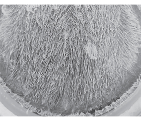
Актуальність. Сучасним актуальним завданням клінічної офтальмології є пошук простих скринінгових та водночас об’єктивних методів обстеження та діагностики патології органа зору. Серед таких діагностичних тестів особливе місце посідає кристалографія, в основі якої — властивість біологічних рідин до кристалоутворення. Серед важливих переваг кристалографії: простота, доступність, можливість динамічного спостереження за розвитком патологічного процесу та ідентифікація змін під час розвитку патологічного процесу. Було доведено, що слізна рідина містить низку компонентів, які реагують на зовнішні впливи та захворювання, що має діагностичне значення. Водночас важливим є отримання узагальненої інформації щодо змін біохімічного вмісту слізної рідини. Тому в пошуках такого підходу ми звернули увагу на тезіографічний метод дослідження біологічних рідин тіла людини, який ґрунтується на тому, що додавання до кристалоутворюючої речовини будь-якої біологічної субстанції призводить до змін у звичайній кристалізації цієї речовини. Такий мікрокристалічний метод називають тезіографією. Мета дослідження: з’ясування можливості тезіографічного дослідження слізної рідини для подальшого його використання як діагностичного скринінгового методу в офтальмологічній практиці. Матеріали та методи. Нами було вивчено зразки слізної рідини від 27 здорових осіб, вік яких коливався від 25 до 57 років. Забір слізної рідини проводили на фільтрувальну смужку, яку після адсорбції на ній слізної рідини висушували та зберігали до дослідження. Для проведення тезіографічного дослідження проводили екстрагування слізної рідини із фільтрувальної смужки. Як базисну кристалоутворюючу речовину використовували 2% розчин хлорної міді у 96° етиловому спирті, який змішували із отриманим екстрактом. Кристалізацію проводили у термостаті при температурі +60 °С. Отримані кристалографічні рисунки фотографували і вивчали візуально та після комп’ютерного збільшення їх розміру, проводячи їх морфологічний опис. Результати. Проведені нами тезіографічні дослідження дозволили з’ясувати кристаломорфологічну характеристику слізної рідини у здорових осіб. Кристалограма базисної слізної рідини здорових осіб представлена у вигляді кристалографічного рисунка, який формується на кристалографічному полі знизу вверх від центрів кристалізації у вигляді первинних фацій, що складаються із довгих дендритів першого порядку з їх віялоподібним розходженням догори з подальшим віялоподібним розгалуженням та утворенням колоскоподібних дендритів з проспективним ростом вгору. На їх окремих ділянках наявні зірчастоподібні фації. При більш детальному вивченні інтегрального кристаломорфологічного рисунка базальної слізної рідини можливо виявити розмежування інтегрального рисунка на 4 морфологічних поля. Формування кристалографічного рисунка біологічного субстрату пов’язано із впливом його компонентів на кристалізаційну решітку самої базисної кристалоутворюючої речовини — хлорної міді. Причому для отримання кристалографічного рисунка достатньо міліграмів слізної рідини, що відповідає половині довжини її адсорбції на фільтрувальній смужці. Висновки. Отримані нами результати дозволили встановити, що навіть незначна кількість слізної рідини, яка адсорбувалася на фільтрувальній смужці, має здатність до тезіографічного кристалоутворення, що вказує на значну чутливість цього методу дослідження. Кристалографічний рисунок слізної базальної рідини можливо отримати із висушених фільтрувальних смужок, що обумовлює відтермінування її дослідження. Зважаючи на простоту тезіографічного методу та його наочність, отримання кристалографічної картини слізної рідини є перспективним скринінговим методом для клінічної офтальмології. При з’ясуванні особливостей кристалограми слізної рідини необхідно вивчати її морфологію після збільшення розміру кристалографічного рисунка.
Background. The modern current task of clinical ophthalmology is the search for simple screening and at the same time objective methods for examination and diagnosis of the ocular pathology. Crystallography, which is based on the properties of biological fluids before crystal formation, has a special place among such diagnostic tests. The important advantages of crystallography are as follows: simplicity, accessibility, possibility of dynamic observation of the pathological process and identification of changes during its development. Tear fluid has been proved to contain a number of components, which respond to external influences and diseases that have diagnostic value. At the same time, it is important to obtain generalized information about changes in the biochemical content of tear fluid. Therefore, in search of such an approach, we turned our attention to the thesiographic method of studying biological fluids of the human body, which is based on the fact that the addition of any biological substance to a crystal-forming material leads to changes in the normal crystallization of this material. This microcrystalline method is called thesiography. The purpose of the research was to clarify the possibility of the thesiographic study of tear fluid for its further use as a diagnostic screening method in ophthalmic practice. Materials and methods. We studied tear fluid samples from 27 healthy individuals, whose age ranged from 25 to 57 years. Tear fluid was collected on a filter strip, which, after tear fluid adsorbed on it, was desiccated and stored until the study. Tear fluid was extracted from the filter paper for the thesiographic study. As a basic crystal-forming material, a 2% solution of copper chloride in 96° ethyl alcohol was used, which was mixed with the obtained extract. Crystallization was carried out in a thermostat at a temperature of +60 °С. The obtained crystallographic patterns were photographed and studied visually and after computer magnification of their size, carrying out their morphological description. Results. Thesiographic studies allowed us to find out the crystal and morphological characteristics of tear fluid in healthy individuals. The crystallogram of the basal tear fluid of healthy individuals is represented as a crystallographic pattern, which is formed on the crystallographic field from the bottom to top from the centers of crystallization as primary facies consisting of long dendrites of the first order with their fern-like divergence to the top with further fern-like branching and the formation of cone-like dendrites with prospective upward growth. There are stellate facies in their separate areas. With a more detailed study of the integral crystal and morphological pattern of the basal tear fluid, it is possible to identify the division of the integral pattern into 4 morphological fields. The formation of the crystallographic pattern of the biological substrate is related to the influence of its components on the crystallization grid of the most basic crystal-forming substance — copper chloride. Moreover, to obtain a crystallographic pattern, milligrams of tear fluid, which corresponds to half the length of its adsorption on the filter paper, are sufficient. Conclusions. The results we obtained allowed us to reveal that even a small amount of tear fluid adsorbed on the filter strip has the ability for thesiographic crystal formation, which indicates the significant sensitivity of this research method. A crystallographic pattern of the tear basal fluid can be obtained from desiccated filter strips, which makes it possible to study it in a delayed manner. Considering the simplicity of the thesiographic method and its clarity, obtaining a crystallographic pattern of tear fluid is a promising screening method for clinical ophthalmology. When clarifying the features of the crystallogram of tear fluid, it is necessary to study its morphology after magnifying the size of the crystallographic pattern.
слізна рідина; тезіографія; кристалографічний рисунок; офтальмологія
tear fluid; thesiography; crystallographic pattern; ophthalmology
Для ознакомления с полным содержанием статьи необходимо оформить подписку на журнал.
- López Solís R., Traipe Castro L., Salinas Toro D., Srur M., Toledo Araya H. Microdesiccates produced from normal human tears display four distinctive morphological components. Biol. Res. 2013. 46. 299-305.
- Traipe-Castro L., Salinas-Toro D., López D., Zanolli M., Srur M., Valenzuela F., Cáceres A., Toledo-Araya H., López-Solís R. Dynamics of tear fluid desiccation on a glass surface: a contribution to tear quality assessment. Biological Research. 2014. 47(25). http://www.biolres.com/content/47/1/25.
- Fedorova O.A. Mozhlyvosti vykorystannia krystalografichnoi harakterystyky ekstrartiv z vnutrishnich organiv trupiv dlia vstanovlennia davnosti nastannia smerti. Sudovo-medychna ekspertyza. 2010. 2. 36-40 [in Ukrainian].
- Biochemistry, Tear Film — StatPearls — NCBI Bookshelf (nih.gov). PMID: 34283502.
- von Thun Und Hohenstein-Blaul N., Funke S., Grus F.H. Tears as a source of biomarkers for ocular and systemic diseases. Experimental eye research. 2013. Dec. 117. 126-137. Doi: 10.1016j.exer.2013.07.015. Epub 2013 Jul 20. PubMed PMID: 23880526.
- Tiffani J.M. Tears in health and disease. Eye. 2003. 17. 923-926.
- Dartt D.A., Willcox M.D. Complexity of the tear film: importance in homeostasis and dysfunction during disease. Experimental eye research. 2013 Dec. 117. 1-3. doi: 10.1016/j.exer.2013.10.008. Epub PubMed PMID: 7034254.
- Van Haeringen N.J. Clinical biochemistry of tears. Survey of Оphthalmology. 1981. 26 (2). 84-96. PubMed PMID: 7034254.
- Bookshelf ID: NBK572136. Biochemistry, Tear Film. PMID: 34283502.
- The Composition of Tears and Their Role in Eye Health. https://www.verywellhealth.com/what-are-tears-made-of-3421862.
- What are Tears Made of? The Biochemistry of Emotion (news-medical.net).
- Simin Masoudi. Biochemistry of human tear film: A review. Exp. Eye. Res. 2022 Jul. 220. 109101. doi: 10.1016/j.exer.2022.109101. Epub 2022 May 1. PMID: 35508212.
- WDH Gillan. Tear biochemistry: a review. The South African Optometrist. 2010. 69 (2). 100-106.
- Zhang X., De Paiva C.S., Su Z., Volpe E.A., Li De-Quan, Pflugfelder S.C. Topical interferon-gamma neutralization prevents conjunctival goblet cell loss in experimental murine dry eye. Experimental Eye Research. 2014. 118. 117-124. ISSN 00144835. doi: 10.1016/j.exer.2013.11.011.
- Kahl J., Busscher N., Doesburg P., Mergardt G., Will F., Schulzova V., Hajslova J., Ploeger A. Application of crystallization with additives to cloudy and clear apple juice. Food Anal. Method. 2017. 10. 247-255.
- Mykhailychenko B.V., Tereshchenko V.P. Crystallographic Portrait of Water. Journal of Water Chemistry & Technology. 2021. 43 (4). 277-280. Dоі: 10.3103/S1063455X21040081.