Вступ
Розриви сухожилків ротаторної манжети плеча (РМП) часто супроводжуються обмеженням як активних, так і пасивних рухів у плечовому суглобі — конт-рактурою плечового суглоба (вторинним адгезивним капсулітом, або secondary stiff shoulder) та змінами щільності кісткової тканини головки плечової кістки [1–4]. Давно відомо про «плямистий остеопороз» головки плечової кістки, що часто супроводжує ідіопатичний адгезивний капсуліт [4, 5]. Аналогічну ситуацію з щільністю кісткової тканини головки плечової кістки ми часто спостерігаємо і під час різноманітних ушкоджень ділянки плечового суглоба з розвитком вторинного адгезивного капсуліту.
Остеопороз є синдромом, що розвивається в результаті адаптивної перебудови формування кісткової тканини у відповідь на метаболічні зміни будь-якої етіології, що відбуваються в організмі [4, 6, 17]. Остеопорозу передує розвиток остеопенії, для якої є характерним зниження мінеральної щільності кісткової тканини від 1 до 2,5 SD (стандартне відхилення) [6, 18].
У літературі зустрічається термін «локальна гіподинамічна резорбція» [19], що найточніше пояснює патогенетичний механізм розвитку явищ локального остеопорозу в кістках скелета за умов низької активності певних груп м’язів. Постійне статодинамічне м’язове навантаження на кістку збільшує її механічну міцність за рахунок деформації кісткового матриксу. Таким чином відбуваються стимуляція надходження мінеральних речовин та активація остеогенезу в головці плечової кістки під дією м’язів РМП, що активно працюють.
Для діагностики остеопорозу золотим стандартом є рентгенденситометрія DEXA (подвійна енергетична абсорбціометрія) [7–9, 16]. Подвійна енергетична рентгенівська абсорціометрія дозволяє визначити вміст солей кальцію, жиру та м’язів в будь-якій ділянці нашого організму. Таким чином, маємо можливість визначити щільність кісткової тканини у будь-якій ділянці головки плечової кістки [7, 10].
Планування оперативного втручання повинно враховувати кількість та характеристики анкерів, що є важливим економічним аспектом будь-якого оперативного втручання. Щільність кісткової тканини головки плечової кістки є показником, що суттєво впливає на кількість та якість необхідних для оперативного втручання анкерів [7, 11]. Чим вища щільність кісткової тканини, тим міцність встановлення анкерів більша, тому кількість використаних фіксаторів може бути меншою. При зниженні щільності кісткової тканини відповідно зменшується міцність фіксації в кістці кожного з встановлених анкерів, що може призвести до міграції анкерів, необхідності застосування великої кількості анкерів або повторної операції [7, 12, 13]. Наявність великих кіст у ділянці великого горбка плечової кістки взагалі унеможливлює постановку фіксаторів і потребує застосування альтернативних варіантів фіксації сухожилків РМП.
Більшість лікувальних установ нашої держави, виконуючи оперативні втручання при контрактурах плечового суглоба різної етіології, не має можливості виконання рентгенденситометрії, що може стати причиною різноманітних ускладнень та незадовільного результату лікування.
Мета дослідження: визначити вплив вторинного адгезивного капсуліту у хворих із розривом сухожилка надостьового м’яза на стан мінеральної щільності кісткової тканини головки плечової кістки.
Матеріали та методи
Обстеження хворих здійснено на рентгеностеоденситометрі Lunar iDXA ME+200082 фірми GE Healthcare (Сінгапур) у стандартних передньозадніх проєкціях у фізіологічному положенні для плечового суглоба та боковій проєкції для поперекового відділу хребта. Обробка даних проводилась на ліцензійному програмному забезпеченні, що постачається в комплекті з рентгеностеоденситометром. Обладнання та програмне забезпечення обслуговуються згідно з технічними нормативами фірми-виробника і метрологічної перевірки не потребують.
Метод дозволяє об’єктивно визначити проникність ділянки скелета для рентгенівського випромінювання й обчислити мінеральну щільність кістки на одиницю площі сканованої поверхні. Цей показник відображається в програмному забезпеченні як BMD і визначається у грамах на один сантиметр квадратний (г/см2) [4, 6, 17].
Проведено рентгенденситометричне обстеження 126 плечових суглобів (63 — із розривом сухожилка надостьового м’яза та 63 — контралатеральних — неушкоджених) у 63 хворих із контрактурою плечового суглоба (порушення пасивних рухів у плечовому суглобі) після розриву сухожилка надостьового м’яза, які з листопада 2015 року по січень 2022 року перебували на амбулаторному та стаціонарному лікуванні у відділі реконструктивно-відновної хірургії верхньої кінцівки Державної установи «Інститут травматології та ортопедії НАМН України» (м. Київ) та Івано-Франківській обласній клінічній лікарні. Вік пацієнтів становив від 35 до 50 років (середній вік — 41,2 ± 15,1 року), чоловіків було 34 (54 %), жінок — 29 (46 %). Середній термін від травми до початку лікування — 64,9 ± 27,8 доби.
На отриманих електронних фотовідбитках рентгенограм за допомогою зазначеного програмного забезпечення встановили по 2 стандартні для всіх хворих зони визначення мінеральної щільності кісткової тканини, як на кінцівці з контрактурою плечового суглоба, так і на аналогічних ділянках контралатеральної кінцівки. Зони вимірювання BMD розміщували так: № 1 — великий горбок плечової кістки, № 2 — середина головки плечової кістки. Кожна зона мала квадратну форму і відповідала 0,90 ± 0,15 см2 площі стандартної рентгенограми (рис. 1). Також всім хворим виконувались стандартні рентгенденситометричні обстеження кульшових суглобів та поперекового відділу хребта.
При аналізі отриманих показників мінеральної щільності в різних зонах вимірювання були отримані показники, що відрізнялись у різних пацієнтів. Тому при аналізі отриманих даних визначали особливості розподілу середніх показників BMD із врахуванням середніх стандартних відхилень.
Критерії включення до дослідження були такими: наявність повношарового розриву сухожилка над-остьового м’яза та контрактури плечового суглоба (контрактурою вважали будь-яке обмеження пасивних рухів у плечовому суглобі), відсутність загальних остеопоротичних змін (рис. 2), вік від 35 до 50 років, відсутність іншої патології плечового суглоба, що ми визначали як клінічно, так і за допомогою додаткових методів дослідження (рентгенографія, магнітно-резонансна томографія), виконання стандартного протоколу рентгенденситометричного дослідження одним спеціалістом.
/20.jpg)
Статистичний аналіз. Статистична обробка даних проводилася за допомогою пакета Statistica 12 (StatSoft, США). Для відображення загальної характеристики вихідних параметрів застосовувалися методи описової статистики із зазначенням середнього показника і стандартного відхилення. Для змінних із нормальним розподілом порівняння груп аналіз проводили за допомогою критерію Стьюдента. Умову рівності дисперсій перевіряли за допомогою критерію Левена. З метою визначення статистичної значущості відмінностей між групами для кількісних (із розподілом, відмінним від нормального) і порядкових змінних був використаний критерій Манна ― Уїтні, для якісних ― критерій χ2 і точний критерій Фішера. Порівняння кількісних і порядкових змінних у залежних вибірках проводили за допомогою критерію Вілкоксона.
Результати
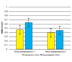
На рис. 3 наведені середні результати показників мінеральної щільності кісткової тканини (BMD) у зонах № 1 ― великий горбок плечової кістки та № 2 ― середина головки плечової кістки кінцівки з конт-рактурою плечового суглоба і розривом сухожилка надостьового м’яза та контралатеральної кінцівки.
/21.jpg)
Як бачимо з рис. 3, у зоні 1 (великий горбок плечової кістки) середня мінеральна щільність кісткової тканини кінцівки з розривом сухожилка надостьового м’яза та контрактурою плечового суглоба становила 0,480 ± 0,265 г/см² і була менше, ніж на здоровій кінцівці, ― 0,638 ± 0,225 г/см² (р = 0,034). Це вказує на вплив розриву сухожилка надостьового м’яза на розвиток остеопоротичних процесів у ділянці великого горбка плечової кістки. У зоні 2 (середина головки плечової кістки) середні показники мінеральної щільності кісткової тканини кінцівки з розривом сухожилка над-остьового м’яза та контрактурою плечового суглоба сягали 0,385 ± 0,118 г/см² і були теж меншими, ніж в аналогічних ділянках контралатеральної кінцівки, ― 0,445 ± 0,148 г/см² (р = 0,021). На нашу думку, даний факт пов’язаний із впливом контрактури плечового суглоба на мінеральний обмін головки плечової кістки та розвитком локальної резорбції кісткової тканини, характерної для вторинного адгезивного капсуліту.
На рис. 4 показана залежність змін мінеральної щільності головки плечової кістки та ділянки великого горбка плечової кістки від кута відведення в плечовому суглобі у хворих із розривом сухожилка надостьового м’яза та привідною контрактурою в плечовому суглобі.
Як видно з риc. 4, ми виявили сильну (r = 0,68; р < 0,01) залежність змін мінеральної щільності кісткової тканини в ділянці великого горбка плечової кістки та слабку, проте вірогідну залежність у ділянці головки плечової кістки (r = 0,44; р < 0,01) від кута відведення в плечовому суглобі у хворих із розривом сухожилка над-остьового м’яза та вторинним адгезивним капсулітом. Даний факт доводить, що розрив сухожилка надостьового м’яза в поєднанні з контрактурою плечового суглоба суттєво впливає на щільність кісткової тканини в ділянці великого горбка плечової кістки.
Обговорення
Ушкодження сухожилків РМП часто призводить до порушення характеру навантаження на великий та малий горбки плечової кістки і, як наслідок, до зменшення щільності кісткової тканини через локальні резорбтивні зміни. Цей факт потребує особливого підходу до вирішення даної проблеми [4, 7, 14, 15]. Наявність контрактури плечового суглоба, як показало наше дослідження, потенціюють дані процеси. Навіть в осіб із нормальними загальними показниками мінеральної щільності кісткової тканини, які взяли участь у нашому дослідженні, ми виявили остеопенію та остеопороз у ділянці головки плечової кістки.
Abtahi та співавт. у своїй статті, присвяченій дослідженню факторів ризику, які впливають на загоєння сухожилків РМП, вказує на наявність остеопоротичних змін у даної групи хворих та на необхідність обов’язкового їх обстеження під час планування оперативного втручання [2]. Вражає той факт, що навіть у такій розвинутій країні, як США, лише 33 % осіб проходять скринінгове дослідження мінеральної щільності кісткової тканини перед виконанням операції на сухожилках РМП і лише 15 % із них в подальшому проходять курс спеціального лікування [8, 9].
Переважна більшість робіт наших західних колег присвячена дослідженню загальних змін мінеральної щільності кісткової тканини після протезування великих суглобів [3], питанням же локальних змін щільності кісткової тканини увага приділяється в поодиноких роботах.
S.W. Chung та співавт. провели унікальне дослідження впливу різних факторів на зрощення сухожилка надостьового м’яза з кісткою після його шва. Була визначена частка незрощень сухожилків РМП на рівні 22,8 %, і доведено, що щільність кісткової тканини має вірогідний вплив на результат шва сухожилків РМП, аналогічний до впливу ступеня ретракції сухожилків РМП, розміру розриву сухожилків РМП та ступеня жирового переродження м’язів РМП [3].
У роботі А.M. Abtahi та співавт. також відзначається, що остеопороз нарівні з цукровим діабетом, курінням, розміром розриву сухожилків РМП та ступенем їх ретракції є негативним фактором для відновлення сухожилків РМП [2]. Однак дана робота, як і багато інших робіт [2, 4, 5, 16], розглядає вплив системного остеопорозу і не розглядає локальні зміни щільності кісткової тканини головки плечової кістки, які, на нашу думку, мають більший вплив на процеси репарації в ділянці плечового суглоба. Саме тому ми виключили з нашого дослідження хворих із системним остеопорозом, що дало можливість більш точно та об’єктивно дослідити вплив контрактури та розриву сухожилка надостьового м’яза на локальні зміни щільності кісткової тканини головки плечової кістки.
Схожим із нашим дослідженням є дослідження J.H. Oh та співавт., в якому автори проводили рентгенденситометричне дослідження обох плечових суглобів у хворих, яким планується виконати шов сухожилків РМП або остеосинтез проксимального епіметафізу плечової кістки [4]. Однак у цьому дослідженні автори не враховували ступінь контрактури плечового суглоба, окрім того, не проведено чіткого розподілу хворих за кількістю ушкоджених сухожилків РМП та віком.
Багато робіт наших західних колег порушують питання не лише діагностики остеопорозу у хворих із розривами сухожилків РМП, а і його лікування як до операції, так і після неї [9, 10, 18]. Аналізуючи отримані нами результати рентгенденситометричного обстеження хворих, ми вважаємо за потрібне застосування у всіх хворих із контрактурою плечового суглоба та розривами сухожилків РМП препаратів, які покращують мінеральну щільність кісткової тканини навіть за відсутності системного остеопорозу.
Сильними сторонами нашого дослідження є виключення з дослідження хворих із системним остеопорозом та остеопенією та віком старше 50 років, що дозволило сформувати однорідну групу хворих, включення до дослідження лише хворих із вторинним адгезивним капсулітом та розривами сухожилка надостьового м’яза (без ушкоджень інших сухожилків РМП), чітко визначені розміри та локалізація зони дослідження, обстеження контралатерального плечового суглоба.
Слабкими сторонами нашого дослідження є: відсутність розподілу групи дослідження за статтю (відомо, що в жінок остеопоротичні процеси перебігають швидше та є більш вираженими), недостатнє програмне забезпечення рентгенденситометра, нехтування термінами захворювання (напевно, що зі збільшенням термінів захворювання зростає і ступінь локального остеопорозу).
Висновки
1. Контрактура плечового суглоба (вторинний адгезивний капсуліт), що виникла в результаті розриву сухожилка надостьового м’яза, призведе до зменшення щільності кісткової тканини головки плечової кістки (р = 0,021) та великого горбка плечової кістки (р = 0,034) навіть у хворих із нормальними загальними показниками мінеральної щільності.
2. Зі зменшенням кута відведення в плечовому суглобі (збільшенням привідної контрактури) у хворих із розривом сухожилка надостьового м’яза та вторинним адгезивним капсулітом зменшується щільність кісткової тканини в ділянці великого горбка плечової кістки (r = 0,68; р < 0,01) та ділянці головки плечової кістки (r = 0,44; р < 0,01).
3. Заходи, спрямовані на хірургічне відновлення цілісності пошкоджених структур РМП, або ліквідація запального процесу з одночасною стимуляцією тканинних репаративних процесів при часткових розривах РМП повинні виконуватися у найкоротші терміни з метою недопущення формування контрактури плечового суглоба.
4. З метою профілактики розвитку явищ локальної резорбції кісткової тканини внаслідок обмеження рухів у плечовому суглобі та гіпотрофії м’язів РМП необхідно приділяти увагу кінезитерапії плечового суглоба з розвитком компенсаторних механізмів збалансованої участі м’язів у забезпеченні мобільності суглоба та необхідного навантаження на кістковий матрикс у пацієнтів із контрактурою плечового суглоба та розривом сухожилка надостьового м’яза.
Конфлікт інтересів. Автори заявляють про відсутність конфлікту інтересів та власної фінансової зацікавленості при підготовці даної статті.
Інформація про внесок кожного автора. Лазарев І.А. — концепція і дизайн дослідження; Богдан С.В. — збирання й обробка матеріалів, аналіз отриманих даних, написання тексту; Юрійчук Л.М. — збирання й обробка матеріалів, аналіз отриманих даних, написання тексту.
Отримано/Received 24.07.2022
Рецензовано/Revised 04.08.2022
Прийнято до друку/Accepted 11.08.2022
Список литературы
1. Mall N.A., Tanaka M.J., Choi L.S., Paletta G.A. Factors affecting rotator cuff healing. J. Bone Joint Surg. Am. 2014. 96. 778-788. DOI: 10.2106/JBJS.M.00583.
2. Abtahi A.M., Granger E.K., Tashjian R.Z. Factors affec-ting healing after arthroscopic rotator cuff repair. World J. Orthop. 2015. 6. 211-220. DOI: 10.5312/wjo.v6.i2.211.
3. Chung S.W., Oh J.H., Gong H.S., Kim J.Y., Kim S.H. Factors affecting rotator cuff healing after arthroscopic repair: Osteoporosis as one of the independent risk factors. Am. J. Sports Med. 2011. 39. 2099-2107. DOI: 10.1177/0363546511415659.
4. Oh J.H., Song B.W., Kim S.H. The measurement of bone mineral density of bilateral proximal humeri using DXA in patients with unilateral rotator cuff tear. Osteoporos. Int. 2014. 25. 2639-2648. DOI: 10.1007/s00198-014-2795-1.
5. Almeida A., Atti V., Agostini D.C., Valin M.R., de Almei-da N.C., Agostini A.P. Comparative analysis on arthroscopic sutures of large and extensive rotator cuff injuries in relation to the degree of osteopenia. Rev. Bras. Ortop. 2015. 50. 83-88. DOI: 10.1016/j.rboe.2015.01.004.
6. Chen X., Giambini H., Ben-Abraham E., An K.N., Nassr A., Zhao C. Effect of bone mineral density on rotator cuff tear: An osteoporotic rabbit model. PLoS One. 2015. 10. DOI: 10.1371/journal.pone.0139384.
7. Denard P.J., Burkhart S.S. Techniques for managing poor quality tissue and bone during arthroscopic rotator cuff repair. Arthroscopy. 2011. 27. 1409-1421. DOI: 10.1016/j.arthro.2011.05.015.
8. Wright N.C., Looker A.C., Saag K.G. The recent prevalence of osteoporosis and low bone mass in the United States based on bone mineral density at the femoral neck or lumbar spine. J. Bone Miner. Res. 2014. 29. 2520-2526. DOI: 10.1002/jbmr.2269.
9. Anderson P.A., Jeray K.J., Lane J.M., Binkley N.C. Bone health optimization: Beyond Own the Bone: AOA critical issues. J. Bone Joint Surg. Am. 2019. 101. 1413-1419. DOI: 10.2106/JBJS.18.01229.
10. Khosla S., Shane E. A crisis in the treatment of osteoporosis. J. Bone Miner. Res. 2016. 31. 1485-1487. DOI: 10.1002/jbmr.2888.
11. Suzuki T., Sukezaki F., Shibuki T., Toyoshima Y., Nagai T., Inagaki K. Teriparatide administration increases periprosthetic bone mineral density after total knee arthroplasty: A prospective study. J. Arthroplasty. 2018. 33. 79-85. DOI: 10.1016/j.arth.2017.07.026.
12. Beaton D.E., Vidmar M., Pitzul K.B. Addition of a fracture risk assessment to a coordinator’s role improved treatment rates within 6 months of screening in a fragility fracture screening program. Osteoporos Int. 2017;28:863–869. DOI: 10.1007/s00198-016-3794-1
13. Spross C., Behrens G., Dietrich T.J. Early arthroscopic repair of acute traumatic massive rotator cuff tears leads to reliable reversal of pseudoparesis: Clinical and radiographic outcome. Arthroscopy. 2019. 35. 343-350. DOI: 10.1016/j.arthro.2018.08.048.
14. Duncan N.S., Booker S.J., Gooding B.W.T., Geoghegan J., Wallace W.A., Manning P.A. Surgery within 6 months of an acute rotator cuff tear significantly improves outcome. J. Shoulder Elbow Surg. 2015. 24. 1876-1880. DOI: 10.1016/j.jse.2015.05.043.
15. Cancienne J.M., Brockmeier S.F., Kew M.E., Deasey M.J., Werner B.C. The association of osteoporosis and bisphosphonate use with revision shoulder surgery after rotator cuff repair. Arthroscopy. 2019. 35. 2314-2320. DOI: 10.1016/j.arthro.2019.03.036.
16. Bernatz J.T., Brooks A.E., Squire M.W., Illgen R.I., Binkley N.C., Anderson P.A. Primary arthroplasty osteoporosis is common and undertreated prior to total joint arthroplasty. J. Arthroplasty. 2019. 34. 1347-1353. DOI: 10.1016/j.arth.2019.03.044.
17. Entezari V., Lazarus M. Surgical considerations in managing osteoporosis, osteopenia, and vitamin D deficiency du-ring arthroscopic rotator cuff repair. Orthop. Clin. North Am. 2019. 50. 233-243. DOI: 10.1016/j.ocl.2018.10.006.
18. Angeline M.E., Ma R., Pascual-Garrido C. Effect of diet-induced vitamin D deficiency on rotator cuff healing in a rat model. Am. J. Sports Med. 2014. 42. 27-34. DOI: 10.1177/0363546513505421.
19. Артеменков А.А. Локальная гиподинамическая остеорезорбция: медикосоциальные причины и патогенетические механизмы. International Journal of Humanities and Natural Sciences. 2021. 56. 144-150. DOI: 10.24412/2500-1000-2021-5-1-144-150.

/19.jpg)
/20.jpg)
/21_2.jpg)