Вступ
Набутий стеноз спинномозкового каналу (ССК) — найбільш поширений прогресуючий хронічний патологічний стан, який характерний для дегенеративних змін у хребті. З віком частота зустрічальності ССК зростає [1–4]. На шийному рівні немієлопатична компресія спинного мозку реєструється у чверті здорового населення [5].
Вимагають уточнення можливі предиктори прогресування ССК з подальшим розвитком неврологічного дефіциту [5]. При цьому клінічно шийна мієлопатія є яскравим проявом ССК, особливо на тлі дегенеративних пошкоджень хребта [5], а провідною клінічною ознакою виявляється больовий синдром, що супроводжує ССК в усіх періодах його розвитку та прогресування. Частота хронічних цервікалгій неухильно зростає [6], і вони є четвертою за частотою причиною непрацездатності [6, 7]. Незважаючи на відмінності будови опорно-рухового апарату людей та тварин [7], у останніх також реєструється больовий синдром у ділянці шийного відділу хребта (ШВХ), що потребує подальшого вивчення.
На сьогодні є небагато інформації про виникнення клінічної симптоматики при ССК в осіб молодого віку. Аналогічно необхідно вивчати відповідний віковий діапазон у тварин за допомогою методів порівняння віку людей та тварин [8, 9].
Стенозованим вважається цервікальний хребетний канал у дорослого з передньозаднім розміром, за різними даними, менше ніж 14–12 мм [10]. Завдяки розвитку методів нейровізуалізації з’явилися ефективні неінвазивні діагностичні критерії щодо діагностики ССК у живої людини [10]. При цьому було виявлено, що анатомічні показники ССК можуть не збігатися з клінічними його проявами у людини. Ці обставини змушують акцентувати увагу на морфометричних та клінічних проявах ССК у тварин [11], у яких зустрічаються дегенеративно-дистрофічні зміни у шийному відділи хребта з проявами болю та іншими симптомами шийної мієлопатії [11, 12].
Таким чином, вивчення вищеописаної патології у тварин може надати новітню інформацію про патофі-зіологічні механізми розвитку ССК, морфологічні зміни у ШВХ, виявити додаткові фактори ризику такого захворювання. А тварини можуть служити більш-менш адекватною моделлю його перебігу.
Мета роботи: клініко-морфометричний порівняльний аналіз набутого ССК у людей та собак.
Матеріали та методи
Обстежено 65 пацієнтів віком від 20 до 65 років та 19 собак масою більше ніж 20 кг і віком від 1 до 14 років Для діагностики ССК долучали комп’ютерну томографію (КТ) ШВХ. При вимірюванні розмірів хребетного каналу зі значенням 12 мм та нижче підтверджувався ССК, крім цього, застосовувався індекс Павлова — Торга, який у нормі становив 1 [10]. Резервний простір вираховувався за допомогою віднімання сагітального діаметра спинного мозку від сагітального розміру хребетного каналу.
Рандомізація за віком у тварин збігалася із середнім віком людей (відповідно 43,4 ± 6,7 року проти 41,5 ± 5,2 року), тобто відбувався перерахунок віку собак на вік людини [8, 11, 12].
Вираженість болю фіксували за допомогою відповідних візуальних аналогових шкал (ВАШ) для людей або тварин [14–16].
Результати та обговорення
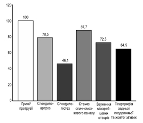
При зверненні пацієнти скаржилися на біль у верхній частині спини, шиї різної інтенсивності та періодичності, оніміння в одній або обох руках, парестезії передпліччя та дистальних відділів верхніх кінцівок. У всіх обстежених на КТ діагностовано: прояви деформуючого спондилоартрозу — 51 пацієнт (78,5 %); звуження міжхребцевих отворів — 47 (72,3 %); деформуючого спондилолістезу — 30 (46,1 %); гіпертрофія поздовжньої і жовтої зв’язок — 42 (64,6 %). Вищенаведене та наявність випинання дисків дорзально або дорзолатерально (100,0 %), кісткових виростів країв хребців та міжхребцевих суглобів сприяли стенотичним змінам спинномозкового каналу та міжхребцевих отворів з розвитком стійкого больового синдрому та неврологічного дефіциту (рис. 1, табл. 1).
/27.jpg)
Важливо, що у більшості пацієнтів виявлено комбінацію таких патоморфологічних дегенеративних змін кістково-хрящового апарату шиї, що компремують вертебральну артерію, ганглії і корінці нервів. Окрім статичних КТ, з метою більш вірогідної діагностики кількості спондилолістезів проводили 3D стандартні постпроцесингові мультипланарні реконструкції.
За даними КТ, ССК на тлі дегенеративно-дистрофічних змін зареєстровано у 57 пацієнтів (87,7 %).
Біль був основною скаргою в усіх пацієнтів. Превалювали цервікалгії — 60 (92,3 %), біль у верхній частині спини реєструвався у 32 (49,2 %) пацієнтів, була характерна іррадіація в верхні кінцівки — 32 (49,2 %). Інтенсивність алгій за ВАШ (табл. 2) коливалася в діапазоні 1–5 балів, середній показник становив 3,1 ± 0,4 бала.
Окрім вищенаведених скарг виявлено: слабкість в одній (14 осіб — 21,5 %) або двох верхніх кінцівках (34 — 52,3 %), тобто в сумі 48 пацієнтів (73,8 %). Крім цього, реєструвалися: м’язові атрофії (14 — 21,5 %), зміни ходи (12 — 18,5 %), слабкість у ногах (13 — 20,0 %), фасцикулярні посмикування (2 — 3,1 %), що наведено в табл. 3.
Моторний дефіцит у вигляді парезів, переважно дистальних відділів рук, зареєстровано у 48 пацієнтів (73,8 %), у ногах пірамідна симптоматика відмічена у 32 (49,2 %) пацієнтів, дисфункція тазових резервуарів — у 7 (10,8 %), а також зустрічалися сенсорні розлади (24 — 36,9 %). Такі прояви шийної мієлопатії повільно прогресували (табл. 4). Пірамідна симптоматика була частішою і більш вираженою у чоловіків.
За даними КТ, дегенеративні зміни хребта максимальними були на рівні хребця С6 з максимальною клінічною кореляцією (неврологічний дефіцит, больовий синдром тощо). Зважаючи на ці обставини, був проведений морфометричний аналіз цього рівня за допомогою індексу Павлова — Торга (< 0,8), який раніше застосовувався для МРТ-досліджень і рентгенологічних знімків. У процесі дослідження виявили вірогідний ССК у людей на КТ-знімках.
Окрім людей обстежено 19 собак масою 20 кг або більше, власники яких вказували на наявність у тварин больового синдрому (100,0 %); зміну поведінки (100,0 %). При обстеженні виявлено порушення ходи (18 — 94,7 %); реакція на пальпацію ділянки шиї (15 — 78,9 %); габітус тварин, які відчувають біль, особливо при рухах шиєю (14 — 73,7 %), напруження тіла (12 — 63,1 %) тощо.
У попередніх наших дослідженнях виявлено, що у великих собак випинання міжхребцевих дисків (р < 0,05) значно більше, ніж у дрібних порід собак, а у котів його майже не буває. Було ретельно досліджено неврологічний та морфометричний стан опорного апарату шиї великих порід собак.
Кісткові вирости країв хребців та міжхребцевих суглобів виявлені майже у всіх собак (18 — 94,7 %) великих порід (р < 0,05) порівняно з іншими (див. наші попередні дослідження). Звуження міжхребцевих отворів, деформація та склеротичні зміни замикальних пластинок, потовщення жовтої та поздовжньої зв’язок зустрічалося у великих порід в три-чотири рази частіше порівняно із собаками масою менше ніж 20 кг (р < 0,01), а у котів були відсутніми. ССК виявлено у 15 (78,9 %) собак (рис. 2). Прояви стенотичних змін розцінено як важливий чинник дегенеративно-дистрофічного процесу, і, ймовірно, саме така органічна патологія зумовлює стійкий больовий синдром.
Як і у випадку людей, больовий синдром фіксували також у тварин, причому інтенсивність болю та поведінка, стан та габітус, згідно з п’ятибальною ВАШ для собак, відповідали стану алгій у людини.
Ущільнення замикальних пластинок, їх деформація та ознаки склеротичних змін виявлено в усіх обстежених собак великих порід.
Візуально та при об’єктивному обстеженні тварин виявлені зміни габітусу, ходи, різного виду та інтенсивності кульгання, пригнічена поведінка, больова реакція на пальпацію.
Аналіз динаміко-статичних даних у собак показав, що незручність при вставанні з положення сидячи або лежачи відзначалася у 10 (52,6 %) обстежених великих собак; зміна статолокомоторики — у 8 (42,1 %); розлади тазових резервуарів — у 3 (15,8 %). У 15 (78,9 %) виявлявся локальний больовий синдром при пальпації ШВХ, ймовірно пов’язаний із стенотичними змінами, корінцевими синдромом (рис. 5).
Ознаки ураження спинного мозку спостерігалися за наявності стенозу у 15 обстежених (78,9 %). Виявлена (табл. 6) легка слабкість однієї (12 — 63,1 % обстежених) чи обох (3 — 15,8 %) передніх кінцівкок, однієї та обох задніх кінцівкок (4 собаки — 21,0 %). У низці випадків можна було почути шаркання пазурів по асфальту — 10 тварин (52,6 %).
При морфометрії ширини спинномозкового каналу у тварин та розрахунках за допомогою індексу Павлова — Торга найчастіше (78,9 %) стенотична патологія зустрічалася на вершині фізіологічного лордозу шиї (С6), аналогічно людям. При дегенеративних змінах хребта на КТ-сканах та сегментарному неврологічному дефіциті клінічно, за наявності больового синдрому, визначено, що у тварин найбільше КТ-проявів мав саме рівень С6 (табл. 7).
За видовими показниками найбільшу частку стенотичних змін спинномозкового каналу ШВХ за індексом стенозу Павлова — Торга виявлено у собак великих порід (78,9 %), що абсолютно збігалося з клінічною картиною.
Чим більша маса тварини, тим пропорційно більший вентро-дорзальний розмір хребця порівняно із сагітальним діаметром спинномозкового каналу.
Хоча спосіб життя тварин відрізняється від способу життя людини і вони мають анатомічні та фізіологічні відмінності, також вони мають схожість патофізіологічних процесів. Таким чином, у нашому дослідженні було відзначено, що на больовий синдром впливають дегенеративні, стенотичні зміни, які мають місце в ШВХ. Усі ці чинники взаємодіють між собою, впливають на структурні, ішемічні, клінічні та інші прояви, прискорюють старіння. Стенотичні, органічні, патофізіологічні та патоморфологічні зміни ШВХ обумовлюють дорсалгії, неврологічні симптоми погіршують якість життя людей. Якщо вважати, що дегенеративно-дистрофічний процес ШВХ може бути предиктором передчасного старіння людей молодого та середнього віку, то вивчення нових етіологічних, патогенетичних механізмів та використання собак великих порід як моделей можуть стати найбільш актуальними з огляду на те, що вік собаки і вік людини піддається перерахунку, а це значить, що фізіологічні процеси у собак проходять у рази скоріше. При такому моделюванні результати нових способів та методів лікування людей, з великою ймовірністю, можуть бути дуже перспективними та корисними для вивчення етіології та патофізіологічних механізмів болі, що, без сумніву, покращить життя людей.
Висновки
За результатами порівняльного аналізу морфометричних даних опорно-рухового апарату ШВХ виявлено таке.
Дегенеративно-дистрофічні зміни реєструвалися у людей вже у молодому і середньому віці, а також в аналогічному віковому діапазоні у домашніх тварин.
Набутий ССК супроводжує дегенеративно-дистрофічні зміни в шиї як у людей (р < 0,05), так і у собак великих порід (р < 0,05).
При цьому розвиваються клінічні прояви шийної мієлопатії (р < 0,05) з переважним стійким больовим синдромом в обстежених людей (р < 0,05) та тварин (р < 0,05).
Максимально часто стенотичні зміни реєструвалися (р < 0,05) на рівні тіла хребця С6 у людей та собак великих порід.
За морфометричними показниками — індексом стенозу Павлова — Торга вони вірогідно збігалися з клінічними проявами: 87,7 % (р < 0,05) у людей та 78,9 % (р < 0,05) у собак великих порід.
Виявлено, що для моделювання дегенеративно-дистрофічних змін в ШВХ треба залучати собак великих порід масою більше 20 кг. При цьому можна значно скоротити час експериментального дослідження та витрати на нього коштів, а також адекватно екстраполювати отримані дані на людей.
Упровадження методів КТ-дослідження, у тому числі морфометричних індексів, у ветеринарну практику ефективне, також економічно вигідне та має максимально гуманний підхід.
Подальші дослідження на моделі ССК на тлі дегенеративно-дистрофічних процесів у собак великих порід дозволять прогнозувати перебіг та прогресування пошкодження ШВХ, адже фізіологічні та патофізіологічні процеси у собак у середньому в 7 раз швидші, ніж у людей.
Наші попередні дослідження вказують, що ступінь вираженості процесу, клініка та морфометричні дані ШВХ тварин залежать від виду, породи, маси, віку, а спільним фактором розвитку дегенеративно-дистрофічних змін у людей та деяких груп тварин може бути старіння організму в цілому і хребта –зокрема.
Конфлікт інтересів. Автори заявляють про відсутність конфлікту інтересів та власної фінансової зацікавленості при підготовці даної статті.
Отримано/Received 05.07.2022
Рецензовано/Revised 22.07.2022
Прийнято до друку/Accepted 01.08.2022
Список литературы
1. Liev A.A. Vertebronevrologiya: stanovlenie, problemyi, perspektivyi. Mіzhnarodniy nevrologіchniy zhurnal. 2009. 25 (3). 12-18 [In Russian].
2. Kozelkin A.A., Kozelkina S.A. Sovremennyie aspektyi diagnostiki i lecheniya vertebrogennyih bolevyih sindromov. Mіzhnarodniy nevrologіchniy zhurnal. 2006. 1 (5). 67-72.
3. Meyer F., Börm W., Thomé C. Degenerative cervical spinal stenosis: current strategies in diagnosis and treatment. Dtsch Arztebl Int. 2008. 105(20). 366-372. doi: 10.3238/arztebl.2008.0366.
4. Lee M.J., Cassinelli E.H., Riew K.D. Prevalence of cervical spine stenosis. Anatomic study in cadavers. J. Bone Joint Surg. Am. 2007 Feb. 89(2). 376-80. doi: 10.2106/JBJS.F.00437. PMID: 17272453.
5. Nouri A., Tessitore E., Molliqaj G., Meling T., Schaller K., Nakashima H., Yukawa Y. et al. Global Spine J. 2022 Feb. 12(1 suppl.). 39S-54S. doi: 10.1177/21925682211036071. PMID: 35174726.
6. Hurwitz E.L., Randhawa K., Yu H., Cote P., Haldeman S. The global spine care initiative: a summary of the global burden of low back and neck pain studies. Eur. Spine J. 2018. 27(Suppl. 6). 796-801.
7. Hautier L., Weisbecker V., Sánchez-Villagra M.R. et al. Skeletal development in sloths and the evolution of mammalian vertebral patterning. Proc. Nat. Acad. Sci. USA. 2010. 107 (44). 18903-18908. doi: 10.1073/pnas.1010335107.
8. Yates K. The Math of Life and Death: 7 Mathematical Principles That Shape Our Lives Paperback. Scribner, April 27, 2021. 288 p.
9. Ulbrich E.J., Schraner C., Boesch C., Hodler J., Busato A., Anderson S.E., Eigenheer S., Zimmermann H., Sturzenegger M. Normative MR. Cervical Spinal Canal Dimensions. Radiology. 2014. 271. 1. 172-182. https://doi.org/10.1148 /radiol. 13120370.
10. Pavlov H., Torg J.S., Robie B., Jahre C. Cervical spinal stenosis. Determination with vertebral body ratio method. Radiology. 1987. 164. 771-5. Cited by: Tierney R.T., Maldjian C., Mattacola C.G., Straub S.J., Sitler M.R. Cervical spine stenosis measures in normal subjects. J. Athl. Train. 2002. 37. 190-3.
11. Veterian Key, Fastest Veterinary Medicine Insight Engine. Chapter 53. Intervertebral Disk Disease. John H. Rossmeisl, Jr. https://veteriankey.com/ intervertebral-disk-disease.
12. Veterian Key, Fastest Veterinary Medicine Insight Engine. Chapter 53. Intervertebral Disk Disease. John H. Rossmeisl, Jr. https://veteriankey.com/ intervertebral-disk-disease/#bib3.
13. Tjahjadi D., Onibala M.Z. Torg ratios based on cervical la-teral plain films in normal subjects. Univ. Med. 2010. 29. 8-10.
14. Stoyanov A.N., Vastyanov R.S., Skorobreha V.Z. Patofiziologicheskie mehanizmyi neyrovegetologii boli. Uch. posobie. Odessa: Astro-print, 2015. 108 [In Russian].
15. Benato L., Murrell J., Knowles T.G., Rooney N.J. Development of the Bristol Rabbit Pain Scale (BRPS): A multidimensional composite pain scale specific to rabbits (Oryctolagus cuniculus). PLoS ONE. 2021. 16(6). e0252417. https://doi.org/10.1371/journal.pone.0252417.
16. Mathews K., Kronen P.W., Lascelles D., Nolan A., Robertson S., Steagall P.V., Wright B., Yamashita K. Guidelines for recognition, assessment and treatment of pain: WSAVA Global Pain Council members and co-authors of this document: J. Small Anim. Pract. 2014 Jun. 55(6). E10-68. doi: 10.1111/jsap.12200.

/27.jpg)
/27_2.jpg)
/28.jpg)
/29.jpg)
/29_2.jpg)