Вступ
За даними Центру громадського здоров’я України, станом на 19 грудня 2021 року в Україні офіційно підтверджено 2536 нових випадків зараження COVID-19, з них 250 дітей, 26 медичних працівників. При цьому у міру накопичення клінічного досвіду терапії SARS-CoV-2 все більше з’являється робіт, у яких висвітлено нові дані про прояви цього вірусного захворювання та наслідки, які можуть впливати як на зміну його клінічної картини, так і на якість життя перехворілого на COVID-19.
Мета роботи: узагальнення результатів літературного пошуку та власного досвіду інтенсивної терапії ендотеліальної дисфункції при коронавірусній інфекції.
Матеріали та методи
В основу роботи покладено результати, отримані за допомогою пошукових інтернет-систем «Google» та «PubMed» за ключовими словами: «інтенсивна терапія SARS-CoV-2», «патофізіологічні зміни при коронавірусній інфекції», «ендотеліальна дисфункція».
Результати
Накопичений власний досвід лікування вкрай тяжких хворих з COVID-19 і результати патогістологічних досліджень хворих, які померли від COVID-19, дозволили визначити 3 основні ланки патогенезу COVID-19, які тісно пов’язані і взаємно доповнюють одна одну:
— Перша — інфікування вірусом епітеліальних клітин слизових оболонок носоглотки, верхніх дихальних шляхів, кишечника, клітин альвеолярного епітелію, імунокомпетентних клітин неспецифічного імунного захисту (нейтрофільних гранулоцитів (НГ), дендритних клітин, моноцитів/мононуклеарних макрофагів, лімфоцитів).
— Друга — гіперпродукція інфікованими клітинами неспецифічного імунного захисту прозапальних цитокінів, у першу чергу ФНП-альфа, IL-6.
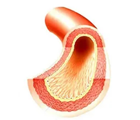
— Третя — вірусне інфікування ендотелію мікросудин з перебудовою внутрішньоклітинного метаболізму на прозапальний лад з розвитком ендотеліту, ушкодженням ендотелію, відщепленням від нього тромбомодуліну і вивільненням у системну циркуляцію тканинного фактора (тканинного тромбопластину) із запуском внутрішньосудинного згортання крові з тромбуванням мікросудин практично всіх органів організму на фоні виснаження системи антикоагуляції і пригнічення фібринолізу.
Досвід зарубіжних дослідників і власний досвід свідчать, що на пізніх стадіях захворювання вирішального значення для позитивного кінцевого результату набуває лікування гіперкоагулопатії, яка розвивається після безпосереднього ураження коронавірусом ендотеліальних клітин судин малого і середнього калібру, а також капілярів практично всіх органів організму.
Тяжкі ендотеліальні ушкодження відбуваються в результаті проникнення вірусу в ендотеліоцити з пошкодженням клітинної мембрани. Розвивається мікроангіопатія, поширений тромбоз судин у результаті вивільнення тканинного фактора із субендотеліоцитарного простору і запуску процесу згортання крові зовнішнім шляхом [1].
Ураження коронавірусом ендотелію супроводжується: активацією коагуляції за рахунок вивільнення тканинного фактора із субендотеліоцитарного простору в судинне русло з активацією VІІ фактора згортання крові і запуском внутрішньосудинного згортання крові, так званого зовнішнього шляху з внутрішньосудинним випаданням тромбіну; пригніченням антикоагулянтної системи, фібринолізу шляхом стимуляції продукції природних інактиваторів фібринолізу — інгібітору активатора плазміногену тканинного типу (PAI-1), інгібітору фібринолізу, активованого тромбіном (TAFI); активацією калікреїн-кінінової системи, активацією тромбоцитів і системи комплементу.
Гістологічно було зареєсроване дифузне ушкодження альвеол: гіалінові мембрани, фібринозний тромбоз судин легень, пошкодження альвеолярного епітелію, тромбози дрібних гілок легеневої артерії, тромбоцитарні і фібринозні тромбози мікросудин [2].
У разі COVID-19 нейтрофільні гранулоцити сильно активуються і зростає їх схильність до агрегації. Активація нейтрофільних гранулоцитів відбувається шляхом експресії на їхній поверхні адгезивних молекул (наприклад, L-селектину), які створюють так званий rolling-пул нейтрофілів (нейтрофіли, що крутяться, обертаються, завихрюються), потім відбувається їх адгезія до ендотелію і вихід із судинного русла в міжклітинний простір, де вони здійснюють хемотаксис в уражені запаленням тканини і виконують роль фагоцитів. На НГ, які не покинули судинне русло, можуть нашаровуватись тромбоцити, фібриноген з тромбуванням мікросудин лейкоцитарно-тромбоцитарними тромбами [3]. Лабораторно це визначається високим умістом незрілих форм нейтрофільних гранулоцитів, низьким рівнем L-селектину, високим процентом клітинної агрегації білих кров’яних тілець, високим рівнем продуктів розпаду нейтрофільних гранулоцитів, у тому числі гістону Н3 [4].
Було виявлене виражене ураження ендотелію легеневих судин: просвіт легеневих судин перекритий агрегатами нейтрофільних гранулоцитів. На електронних сканограмах були зареєстровані пошкодження мікросудин легень, опорний, стовбуровий, інвагінуючий ангіогенез, ураження ендотелію легеневих артерій малого і середнього калібру, капілярів [5].
На сьогодні вважається, що першим кроком до розуміння патогенезу COVID-19 є інформація про те, що ключове значення в процесі інфікування ендотеліоцитів мають рецептори ангіотензинперетворюючого гормона/фактора (АПФ), які розташовуються на їхній поверхні.
Після взаємодії з рецептором АПФ2 на поверхні ендотеліоцитів коронавірус проникає всередину ендотеліоцита і в кінцевому результаті необоротно пошкоджує його із вивільненням у кровотік із субендотеліоцитарного простору тканинного фактора (тканинного тромбопластину), який активує VІІ фактор згортання крові із запуском каскаду системи згортання крові з перетворенням протромбіну в тромбін і фібриногену у фібрин. Одночасно продукти розпаду вірусів і нейтрофільних гранулоцитів активують систему комплементу, яка активується ХІІа фактором згортання крові, який за допомогою компонентів комплементу активує коагуляційний каскад.
Як відомо, АПФ перетворює ангіотензин І в ангіотензин ІІ (АТ-ІІ) і являє собою неспецифічну гідролазу, яка також інактивує брадикінін. Тому застосування інгібіторів АПФ (на сьогодні ключової групи гіпотензивних препаратів) супроводжується накопиченням в організмі брадикініну, що у деяких пацієнтів клінічно проявляється нестримним кашлем.
В експериментальних і клінічних дослідженнях було встановлено, що застосування інгібіторів АПФ призводить до відновлення функцій ендотелію, пригнічення проліферації і міграції клітин гладкої мускулатури, нейтрофілів і моноцитів, зниження оксидативного стресу, посилення ендогенного фібринолізу, зниження агрегаційної здатності тромбоцитів, антиатерогенної дії і стабілізації атеросклеротичної бляшки [6].
На сьогодні вважається, що АТ-ІІ є біологічним активатором двох типів рецепторів АПФ — типу І і ІІ. Активація ангіотензином ІІ АПФ-рецепторів І типу на поверхні ендотеліоцитів змінює їхній метаболізм на прозапальний лад з протромботичною, прооксидантною, профібротичною перебудовою і вазоконстрикцією. Активація ангіотензином ІІ АПФ-рецепторів ІІ типу на поверхні ендотеліоцитів справляє протилежний вплив: протизапальний, антитромботичний, антиоксидантний, антифібротичний, вазодилатуючий. У разі COVID-19 блокуються ангіотензин-ІІ-активовані АПФ-рецептори ІІ типу. Накопичення в організмі ангіотензину ІІ зумовлює активацію тромбоцитів, системне утворення тромбіну, пошкодження ендотелію, недостатність антикоагуляції, тромбози, гіперфібриноліз, гіперфібриногеноліз, прооксидантний ефект, фіброз, вазоконстрикцію (вазоконстрикторний ефект ангіотензину ІІ у 20 разів перевищує вазоконстрикторний ефект норадреналіну). У разі COVID-19, накопичення ангіотензину ІІ призводить до активації рецепторів ангіотензинперетворюючого гормона І типу. Виділяють три основних ефекти цієї активації: ангіотензин-ІІ-активовану коагулопатію, гіперфібриноліз і гіперфібриногеноліз, активацію кінін-калікреїнової системи, дисеміноване внутрішньосудинне згортання крові. Ангіотензин ІІ вважається функціональним рецептором для коронавірусів. Ангіотензин ІІ накопичується у високих концентраціях у тканинах і активує АПФ-рецептори І типу епітеліоцитів, клітин неспецифічного імунного захисту й ендотеліоцитів, з якими взаємодіє коронавірус своїм протеїном S i в цьому місці розплавляє клітинну мембрану й інфікує клітину. Уражаються ендотеліоцити судин практично всіх органів: мозку, серця, легень, печінки, нирок, селезінки, шлунка, тонкого кишечника, товстого кишечника, слизових оболонок рота і носа, клітин гладкої мускулатури, клітин альвеолярного епітелію, шкіри, лімфатичних вузлів і кісткового мозку.
Одночасно ХІІа фактор перетворює прекалікреїн у калікреїн, який, у свою чергу, перетворює плазміноген в плазмін. Крім того, ХІІа фактор сам активує плазміноген з перетворенням його в плазмін. Тільця Вейбела — Паладе також активують плазміноген з перетворенням його в плазмін, а також сприяють вивільненню із ендотеліоцитів тканинного активатора плазміногену (tPA).
На поверхні фібрину, який викликав тромбування судин, утворюється потрійний комплекс — плазміноген, тканинний активатор плазміногену, лізинова ділянка фібрину, з утворенням плазміну, який лізує фібрин з утворенням продуктів деградації фібрину і фібриногену, а також D-димера.
Плазмін розщеплює протеїн S коронавірусу в позаклітинному просторі, таким чином підвищуючи його здатність взаємодіяти з рецепторами ангіотензинперетворюючого гормона на мембранних поверхнях епітеліальних і ендотеліальних клітин людини і, ймовірно, полегшуючи інвазію вірусу в клітину. Плазмін протеолітично розщеплює фібрин і фібриноген з підвищенням концентрації D-димера та інших продуктів деградації фібрину і фібриногену в бронхоальвеолярній рідині і плазмі крові [7].
Велике значення в патогенезі COVID-19 має активація системи комплементу. Компонентами комплементу активуються тромбоцити, коагуляція, розвивається недостатність антикоагуляції, пригнічення фібринолізу [8].
Вважається, що активація коагуляції, пригнічення антикоагуляції, пригнічення фібринолізу за рахунок продукції PAI-1 i TAFI спрямовані на обмеження поширення вірусної інвазії і локалізацію вогнищ вірусного запалення.
У результаті вищенаведених патофізіологічних перетворень в організмі хворого, інфікованого COVID-19, розвиваються два різновиди гострого респіраторного дистрес-синдрому (ГРДС) — ГРДС І типу і ГРДС ІІ типу. ГРДС І типу — це синдром, який формується за рахунок активації клітин неспецифічного імунного захисту — нейтрофільних гранулоцитів і моноцитів, має назву «локальний фізіологічний імунотромбоз» і характеризується: системним запаленням, активацією тромбоцитів, активацією системи згортання крові, виснаженням системи антикоагуляції, порушенням фібринолізу, активацією ендотелію з розвитком ендотеліту (перебудовою метаболізму ендотелію на прозапальний лад), який може переходити в ГРДС ІІ типу. ГРДС ІІ типу отримав назву «системний патологічний мікросудинний тромбоз», який характеризується: вираженою системною запальною реакцією з «цитокіновим штормом», активацією тромбоцитів з їх дегрануляцією (вивільненням із них серотоніну, тромбоцитарного тромбопластину/фактора 3 тромбоцитів), їх функціональним виснаженням, агрегацією і споживанням, пошкодженням ендотелію, вивільненням тканинного фактора із субендотеліального простору, системним утворенням тромбіну, порушеннями в системі антикоагуляції, персистуючим пригніченням фібринолізу. У результаті пошкодження ендотелію і тромбування мікросудин у разі COVID-19 уражаються практично всі органи і системи.
Тромбоцитопенія вважається предиктором летального наслідку при COVID-19. У разі вмісту тромбоцитів до 50 × 109/л летальність досягає 92,1 %; від 50 до 100 × 109/л — 61,2 %; від 100 до 150 × 109/л — показник летальності становить 17,5 % [9]. Переважно виживають ті пацієнти, у яких вміст тромбоцитів перевищує 200 × 109/л [10].
Чутливість методу визначення концентрації D-димера становить 92,3 %, специфічність — 83,3 %. У разі концентрації в плазмі D-димера менше ніж 2 мкг/мл показник очікуваного виживання досягає 100 %. У випадках, коли концентрація D-димера перевищує 2 мкг/мл, летальність на 30–40-й день захворювання досягає 80 % [11] (у більшості лабораторій України концентрація D-димера визначається в мкг/л, таким чином, вищевказані показники необхідно множити на 1000).
У перебігу COVID-19 виділяють наступні фази/періоди: інкубаційний період — у середньому 5 діб; період клінічних проявів — 5–11-та доба — характеризується лихоманкою, кашлем, головним болем, діареєю; рання легенева фаза — 11–14-та доба — характеризується гіпоксією середнього ступеня (при інгаляції кисню через носові канюлі з швидкістю 4 л/хв — SaО2 < 94 %); пізня легенева фаза — 14-та — 28-ма доба — характеризується прогресивним наростанням гіпоксії [12].
Гістологічні ознаки COVID-19: поширений тромбоз із мікроангіопатією, тромбоз капілярів альвеоло-капілярної мембрани, який за інтенсивністю в 9 разів перевищує відповідний тромбоз у разі грипозної пневмонії [13].
Морфологічними ознаками ураження органів при COVID-19 вважаються мікротромбози судин легень, печінки, нирок, серця, головного мозку [14].
Частота виникнення гострої тромбоемболії легеневої артерії (ТЕЛА) у хворих на COVID-19 становить у перерахунку на всіх, хто захворів, у середньому 15 % (з коливаннями від 10 до 21 %), а в палатах інтенсивної терапії — у середньому 23 % (з коливаннями від 17 до 32 %) [15, 16].
Порушення параметрів коагуляції вважається фактором негативного прогнозу у пацієнтів з коронавірусною пневмонією. Предикторами невиживання на ранніх стадіях захворювання вважаються: подовження протромбінового часу, високі рівні продуктів деградації фібрину, фібриногену і D-димера, високі концентрації фібриногену, а на пізніх стадіях — подовження протромбінового часу, високі рівні продуктів деградації фібрину, фібриногену і D-димера, високе співвідношення ПДФ та D-димера (> 7), низька концентрація фібриногену, ознаки дисемінованого внутрішньосудинного згортання крові [17].
Профілактика виникнення та лікування коагулопатій і тромбоемболічних ускладнень у хворих на COVID-19
На сьогодні особливості проведення антикоагулянтної терапії у хворих на COVID-19 полягають в такому: усім хворим на COVID-19 призначаються низькомолекулярні гепарини (НМГ) у профілактичних дозах; хворим палат інтенсивної терапії НМГ призначаються у зростаючих дозах; у разі наявності у хворих тромбозу глибоких вен або ТЕЛА НМГ призначаються в лікувальних дозах; хворим з ГРДС НМГ призначаються у зростаючих дозах [18].
Рекомендації Міжнародної асоціації з проблем тромбозу і гемостазу (ISTH)
1. Для профілактики венозних тромбоемболій рекомендується перевагу надавати НМГ або фондапаринуксу.
2. Рекомендуються стандартні профілактичні дози НМГ, але можуть використовуватись і середні терапевтичні дози.
3. У хворих, які знаходяться в критичних станах, тромбопрофілактика може здійснюватись НМГ і нефракціонованим гепарином (НФГ).
4. У хворих з ожирінням доза гепаринів розраховується на актуальну масу тіла або з використанням індексу маси тіла + 50 % розрахункової дози.
5. Після виписки із стаціонару антикоагулянтна терапія повинна бути продовжена протягом від 14 до 30 діб у пацієнтів з факторами ризику тромбоемболічних ускладнень (ТЕУ).
Рекомендації CHEST (настанови і власний досвід авторів)
1. Тромбопрофілактика НМГ або фондапаринусом має переваги над НФГ.
2. Тромбопрофілактика НМГ і НФГ має переваги над механічними методами профілактики.
3. Дози гепаринів для тромбопрофілактики повинні перевищувати середньотерапевтичні дози і повинні збільшуватись пропорційно масі тіла.
4. Не рекомендують застосовувати методи механічної профілактики на фоні гепаринотерапії.
5. Після виписки із стаціонару тромбопрофілактику антикоагулянтами продовжують.
Таким чином, на сьогодні, по суті, не існує єдиного затвердженого міжнародного протоколу проведення антикоагулянтної терапії у пацієнтів з COVID-19 [19].
Антикоагулянтна терапія з метою зниження частоти тромбоемболічних ускладнень
Хворим середнього ризику ТЕУ з ІМТ менше ніж 30 кг/м2, які не потребують і потребують оксигенотерапії, рекомендують призначати НМГ у профілактичних дозах або фондапаринукс:
— еноксапарин 4000 МО п/ш 1 раз на добу або 2000 МО п/ш 1 раз на добу, якщо кліренс креатиніну (Clcr) від 15 до 30 мл/хв;
— беміпарин 3500 МО п/ш 1 раз на добу, якщо Clcr > 30 мл/хв;
— дальтепарин 5000 МО п/ш 1 раз на добу, якщо Clcr > 30 мл/хв;
— фондапаринукс 2,5 мг п/ш 1 раз на добу, якщо Clcr > 50 мл/хв.
Хворим з високим ризиком ТЕУ з ІМТ більше ніж 30 кг/м2 за відсутності інших факторів ризику тромбозу, а також у пацієнтів з ІМТ більше ніж 30 кг/м2 з наявністю інших факторів ризику тромбозу, яким проводиться оксигенація з високим потоком О2 через носові канюлі або проводиться штучна вентиляція:
— еноксапарин 4000 МО п/ш 2 рази на добу;
— еноксапарин 6000 МО п/ш 2 рази на добу, якщо маса тіла більша за 120 кг;
— НФГ 200 МО/кг в/в протягом 24 годин, якщо Clcr < 30 мл/хв.
Моніторинг антикоагулянтної терапії доцільно здійснювати за допомогою моніторингу анти-Ха-факторної активності. Необхідно уникати передозування НМГ. Тому треба підтримувати анти-Ха-факторну активність на рівні нижче за 1,2 МО/мл для еноксапарину. У разі застосування НФГ анти-Ха-факторну активність рекомендують підтримувати на рівні 0,3–0,5 МО/мл (контроль умісту тромбоцитів здійснювати кожні 48 годин) [20].
Хворим дуже високого ризику ТЕУ, у яких мають місце виражена системна запальна реакція (фібриноген більше за 8 г/л), гіперкоагуляція (концентрація D-димера більша за 3 мкг/мл або більша за 3000 мкг/л), яким проводиться ЕКМО або лікування антикоагулянтами тривалої дії, рекомендовано:
— НМГ (еноксапарин) у лікувальних дозах (100 МО/кг п/ш 2 р/добу, не перевищуючи дозу 10 000 МО 2 рази на добу);
— НФГ рекомендують застосовувати в дозі 500 МО/кг протягом 24 годин, якщо кліренс креатиніну нижчий за 30 мл/хв.
Дози антикоагулянтів повинні бути скориговані у хворих із синдромом поліорганної недостатності та з коагулопатією споживання.
Гепаринорезистентність розглядають як недооцінену проблему. Антикоагулянтна активність гепарину залежить від концентрації в плазмі антитромбіну ІІІ. Гепарин зв’язується з АТ ІІІ і активованими факторами згортання крові ІІа, ІХа, Ха, ХІа, ХІІа в потрійний комплекс з наступною інактивацією активованих факторів згортання крові. До гепаринінактивуючих білків відносяться: фактор 4 тромбоцитів, фібриноген, VІІІ фактор згортання крові, гістидинзбагачений глікопротеїн. У пацієнтів з COVID-19, які знаходились у палатах ІТ, резистентність до гепарину зафіксована у 80 % випадків, а у 100 % пацієнтів — зниження активності терапевтичних доз НМГ. Для поповнення запасів АТ ІІІ необхідно переливати нативну плазму — 1 доза на тиждень [21].
Говорячи про ендотеліальну дисфункцію при SARS-CoV-2, не можна не згадати про препарати, що містять L-аргінін. І тут важливо відзначити не лише їх пряму дію, спрямовану на збільшення рівня NO [22]. Справа в тому, що нормальна імунна система залежить від кількості L-аргініну, доступного в організмі. Відомо, що аргіназа є невід’ємною частиною певних субпопуляцій гранулоцитів, які можуть вивільнятися локально або систематично при виникненні імунної відповіді. Крім того, відбувається накопичення незрілих мієлоїдних клітин, які експресують аргіназу, що вивільняється при боротьбі з певними захворюваннями. Ці клітини, що експресують аргіназу, можуть зменшувати кількість L-аргініну [23].
У роботах R. Geiger та співавт. було показано, що функція Т-клітин залежить від рівня L-аргініну [24]. Повідомлялося про зниження здатності лімфоцитів до проліферації у пацієнтів із сепсисом у критичному стані, що корелювало зі зменшенням доступності L-аргініну [25]. Більше того, було виявлено, що введення L-аргініну корисне для підтримки імунного гомеостазу, особливо з точки зору функції Т-клітин та макрофагів [26]. Фактично L-аргінін необхідний для перемикання макрофагів з M1 на M2.
Було показано, що дефіцит L-аргініну призводить до зниження проліферації Т-клітин та викликає зниження відповіді в опосередкованій Т-клітинами пам’яті [27]. Аналізи in vitro підтвердили, що L-аргінін може відновлювати функцію Т-клітин [28]. Механічні імуносупресивні ефекти мієлоїдних супресорних клітин (MDSC) через виснаження L-аргініну та мітохондріальну дисфункцію лімфоцитів були продемонстровані на моделях раку. Збільшення кількості MDSC, яке спостерігається при COVID-19, безпосередньо корелює з підвищеною активністю аргінази та лімфопенією. Моноцитарні MDSC значно збільшилися в крові пацієнтів із COVID-19 і були тісно пов’язані з тяжкістю захворювання. Було показано, що MDSC пригнічують проліферацію Т-клітин і продукцію IFN-γ, принаймні частково, за допомогою аргіназозалежного механізму, що переконливо вказує на роль цих клітин у дизрегульованій імунній відповіді при COVID-19 [29]. Дійсно, MDSC експресують високі рівні аргінази, яка метаболізує L-аргінін до орнітину та сечовини, ефективно виснажуючи цю амінокислоту з мікрооточення. Відомо, що виснаження L-аргініну пригнічує передачу сигналів рецептора Т-клітин, що в кінцевому підсумку призводить до дисфункції Т-клітин і до збільшення генерації активних форм кисню, тим самим загострюючи запалення [30].
У недавньому дослідженні, присвяченому COVID-19, було визначено біодоступність L-аргініну у трьох когортах: безсимптомних здорових дорослих, дорослих, госпіталізованих з COVID-19, та дітей, госпіталізованих з COVID-19, і було встановлено, що як дорослі, так і діти з COVID-19 демонстрували значно нижчі рівні L-аргініну в плазмі (а також біодоступність L-аргініну) порівняно з контрольною групою [31]. Крім того, низьке співвідношення L-аргініну та орнітину, яке спостерігається у пацієнтів з COVID-19, вказує на підвищення активності аргінази у цих пацієнтів. В іншому дослідженні було показано, що рівні L-аргініну в плазмі обернено корелюють з тяжкістю COVID-19 [32]. Це дослідження також показало, що експресія активованого комплексу GPIIb/IIIa (PAC-1), який, як відомо, бере участь в активації тромбоцитів та тромбоемболічних подіях, вища на тромбоцитах пацієнтів з тяжкою формою COVID-19 порівняно зі здоровими людьми контрольної групи та корелює з плазматичною концентрацією L-аргініну [32].
Ці докази суперечать нещодавно запропонованій стратегії виснаження L-аргініну при COVID-19, заснованій на припущенні, що деякі етапи життєвого циклу вірусу SARS-CoV-2 можуть залежати від залишків L-аргініну (наприклад, білок нуклеокапсид містить 6,9 % L-аргініну) [33].
Фактично було показано, що зниження біодоступності L-аргініну викликає зниження відповіді та функції Т-клітин, що зрештою призводить до підвищеної сприйнятливості до інфекцій. Дванадцять тижнів безперервного прийому L-аргініну значно знизили рівень IL-21 [34], у той час як NO, як було показано, пригнічує проліферацію та функцію людських клітин Th17, що відіграє важливу роль у патогенезі «цитокінового шторму» та гіперзапальних явищ, що спостерігаються у пацієнтів із COVID-19.
Більш високі рівні L-аргініну пов’язані з нижчими рівнями CCL-20, ліганду CCR6, що є частиною системи хемотаксису, яка індукується у відповідь на коронавірусні інфекції [35]. Дані факти роблять доцільним використання донаторів L-аргініну у схемі лікування пацієнтів із коронавірусною інфекцією.
Висновки
Проведений огляд переконливо показує, що інфекція SARS-CoV-2 сприяє розвитку ендотеліту у різних органах як наслідок вірусного ураження. Саме наявністю COVID-19-індукованого ендотеліту можна пояснити системне порушення мікроциркуляції у різних судинних руслах та їх клінічні наслідки.
Конфлікт інтересів. Автори заявляють про відсутність конфлікту інтересів та власної фінансової зацікавленості при підготовці даної статті.
Інформація про внесок кожного автора. Бондар М.В. — збір літературного матеріалу, підготовка результатів дослідження до аналізу; Пилипенко М.М. — визначення дизайну дослідження, узагальнення результатів дослідження; Лоскутов О.А. — аналіз матеріалів дослідження, узагальнення результатів дослідження.
Отримано/Received 06.12.2021
Рецензовано/Revised 13.12.2021
Прийнято до друку/Accepted 15.12.2021