Вступ
Визначення точних параметрів розгинання в гомілковостопному суглобі (ГС) залишається важливим завданням. Поряд із традиційною гоніометрією упродовж останнього часу було створено багато інструментів, які використовують для визначення амплітуди рухів у ділянці гомілковостопного суглоба, кожен з яких характеризується різним рівнем технічної складності та вірогідності отриманих результатів. Незважаючи на широкий спектр наявних засобів, серед науковців немає єдиної узгодженої думки щодо переваг використання конкретного методу, що значно ускладнює порівняння клінічних досліджень та застосування в практичній медицині [1–3]. Рентгенографія залишається золотим стандартом (evidence based) вимірювання амплітуди рухів у суглобах [4], однак ця методика має обмежене використання на практиці з огляду на наявність променевого навантаження [5]. Необхідність покращити ефективність вимірювань призвела до пошуків дослідниками нових методів оцінки діапазону розгинання в ГС, які мали б значний рівень надійності та відтворюваності. Так, поряд із традиційною гоніометрією нещодавно були розроблені додатки інклінометра для смартфонів, що робить їх популярним вибором для клініцистів [6–10]. Систематичний огляд, проведений J. Keogh et al. [11], продемонстрував вагомі докази на підтримку використання мобільних інклінометрів для вимірювання рухів у суглобах. Проте, незважаючи на велику кількість публікацій, присвячених визначенню обсягу розгинання у ГС з використанням гоніометрів і мобільних інклінометрів, досліджень, які порівнювали б ці методи з рентгенографічним методом, на жаль, недостатньо, що обумовлює важливість та актуальність подальших пошуків.
Мета роботи — провести порівняльний аналіз валідності гоніометричного, інклінометричного та рентгенологічного методів вимірювання обсягу розгинання у гомілковостопному суглобі.
Матеріали та методи
Матеріалом для роботи стали результати обстеження 25 фізично здорових осіб (50 гомілковостопних суглобів) на базі КНП «Ірпінська центральна міська лікарня». Середній вік пацієнтів становив 25,8 ± 5,2 року; переважали чоловіки — 18, жінок було 7; середнє значення індексу маси тіла — 25,01 ± 5,01. Критерії виключення: наявні неврологічні та вестибулярні захворювання, анамнез травм чи хірургічних втручань на нижніх кінцівках, перенесений у минулому перелом кісток стопи та гомілки. Письмова інформована згода була отримана від усіх учасників дослідження. Вимірювання амплітуди активного розгинання у ГС проводили у положенні навантаження (тест «випаду» — weight-bearing lunge test, WBLT), згідно з рекомендаціями American Medical Association [12, 13]. Оцінку діапазону розгинання у ГС проводили за допомогою універсального двоплощинного гоніометра (діапазон градусної шкали 1°) та інклінометра на основі додатка до смартфона (The Clinometer Smartphone Application™) [14], який використовує внутрішній акселерометр пристрою для визначення просторового положення та вимірювання кутових показників.
Методика дослідження. Обстежуваний знаходився у вертикальному положенні, стопа розташовувалась перпендикулярно до стіни. Учасникам було запропоновано здійснити нахил вперед, згинаючи колінний суглоб обстежуваної кінцівки (паралельно ІІ пальцю стопи) до моменту контакту зі стіною, при цьому п’ятка залишалась на опорній поверхні. Правильне положення заднього відділу стопи контролювалося дослідником. Після досягнення учасником максимального положення «випаду» дослідник проводив вимірювання амплітуди розгинання, використовуючи стандартний двоплощинний гоніометр, проксимальне плече якого було розміщено паралельно діафізу 5-ї плеснової кістки, а дистальне плече розміщувалось паралельно латеральному краю малогомілкової кістки з центром ротації в ділянці латеральної кісточки (рис. 1а). Після реєстрації гоніометричних показників, не змінюючи положення кінцівки, дослідник розміщував інклінометр на передній поверхні гомілки, на 2 см дистальніше горбистості великогомілкової кістки, та фіксував значення максимального показника інклінометра. Перед початком кожного вимірювання цифровий інклінометр був відкалібрований відносно вертикальної площини, з метою забезпечення єдиної узгодженої вихідної точки (рис. 1б).
/27.jpg)
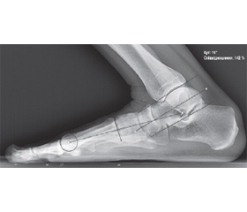
Усім учасникам виконували рентгенографію обох ГС з навантаженням у стандартній медіолатеральній проєкції в положенні максимального розгинання. Латеральний край стопи був позиціонований з боку рентгенологічної касети. На отриманих рентгенограмах вимірювали рентгенологічні (скіалогічні) показники: кут, утворений лініями, що з’єднують діафізарні центри малогомілкової кістки та 5-ї плеснової кістки (малогомілково-5-плесновий кут, Fibula — MT5) (рис. 2); латеральний тало-1-метатарзальний кут (латеральний кут Meary’s) за рекомендованою методологією [15]. Рентгенологічні параметри вимірювали з інтервалами 1° за допомогою програмного забезпечення для редагування рентгенологічних зображень (Vidar Dicom Viewer; ООО «Видар-ИнфоРад», Москва, РФ). Усі вимірювання проводились на кожній кінцівці тричі, середнє значення кожного виміру використовували як репрезентативне значення для аналізу даних.
/28.jpg)
Отримані дані та результати вимірювань заносили в електронні таблиці, застосовували методи описової статистики (середнє, стандартна помилка). Суттєві відмінності визначали за допомогою однофакторного дисперсійного аналізу (ANOVA). У разі виявлення статистично значимих відмінностей між групами додатково проводилося порівняння сукупностей попарно за допомогою апостеріорного критерію Шеффе. Коефіцієнт варіації (CV) був розрахований для визначення відтворюваності вимірювань. Апріорна статистична значимість була встановлена на рівні р ≤ 0,001. Усі розрахунки проводили в Microsoft Office Excel 2016 з використанням наданого пакета програм.
Результати та обговорення
Обмеження розгинання в гомілковостопному суглобі тісно пов’язане з низкою травм і захворювань нижніх кінцівок: плантарним фасціїтом [16], ахілобурситом [17], наслідками переломів кісточок та ушкоджень зв’язкового комплексу надп’ятково-гомілкового та колінного суглобів [18]. Розуміння клініцистами взаємозв’язку між амплітудою розгинання та функціональними характеристиками ГС, точна оцінка та динаміка відновлення розгинання залишаються важливим компонентом визначення ефективності та якості ортопедичної діагностики. Вимірювання діапазону рухів в положенні навантаження отримало широку популярність серед дослідників, оскільки краще відображає функціональні можливості суглоба (ходьба, біг, підйом сходами), має більшу надійність та репрезентативність порівняно з показниками, отриманими в положенні без навантаження [3]. Систематичний огляд, проведений С. Powden et al. [19], показав, що WBLT-тест має високу чутливість та експертну погодженість при оцінці активного розгинання у ГС, що робить його зручним інструментом для клінічних досліджень. Враховуючи дані літературних джерел щодо відсутності суттєвої різниці в амплітуді рухів між правим і лівим ГС [20, 21], для статистичного аналізу в нашому дослідженні використали об’єднану вибірку. За результатами вимірів середнє значення розгинання у ГС, виміряне за допомогою двоплощинного кутоміра, становило 37,62 ± 5,56°; середнє значення інклінометра — 40,61 ± 5,15°, що узгоджується з раніше повідомленими нормативними значеннями для здорових осіб [1, 22]. Середнє значення рентгенологічних параметрів (малогомілково-5-плесновий кут), визначеного на бокових навантажувальних рентгенограмах, становило 23,69 ± 7,25°, що значно менше порівняно з досліджуваними методами (рис. 3).
Розрахунки показали статистично значиму різницю між значеннями малогомілково-5-плеснового кута та показниками інклінометричного і гоніометричного методів вимірювання тильного розгинання в гомілковостопному суглобі (p < 0,001). Між гоніометричним та інклінометричним методами вимірювання статистично значуща різниця виявлена не була (p < 0,001). Коефіцієнт варіації визначали за формулою:
де SD — середнє квадратичне відхилення, x — середнє арифметичне значення.
Отримані значення CV для трьох методів вимірювань показали кращу відтворюваність інклінометрії (0,15) та гоніометрії (0,13) порівняно з рентгенологічним методом (0,31) (p < 0,001). Не було виявлено суттєвої різниці в CV між результатами вимірів у правому та лівому гомілковостопних суглобах (табл. 1).
/29.jpg)
Використання рентгенографії для рутинної оцінки рухів у гомілковостопному суглобі в клінічних умовах не застосовують, проте це дозволяє використовувати її для встановлення обґрунтованості інших методів, таких як гоніометрія чи інклінометрія. Незважаючи на високу відтворюваність гоніометричного та інклінометричного методів вимірювання, недоліком цих засобів залишається висока імовірність виникнення потенціальної помилки, пов’язаної із складнощами позиціонування заднього відділу стопи при обстеженні, що надає рентгенологічному дослідженню додаткову перевагу та інформативність у визначенні істинного діапазону розгинання у ГС. Так, P. Dayton et al. [23], порівнюючи вплив супінованого, нейтрального і пронованого положення заднього відділу стопи на показники розгинання в гомілковостопному суглобі, виявили мінімальну різницю у скіалогічних показниках при кожному з трьох положень стопи із загальною різницею в 0,35°, в той час як клінічне вимірювання розгинання виявило середню різницю в 14° між супінованим і пронованим положенням стопи. Схоже дослідження K. Cady et al. [24] показало, що обсяг розгинання у ГС також суттєво відрізняється залежно від позиції заднього відділу стопи при виконанні WBLT.
/29_2.jpg)
З клінічної точки зору оцінка розгинання у ГС переважно залежить від його мобільності. Незважаючи на це, все більше дослідників акцентують увагу на важливості оцінки впливу суміжних суглобів (колінного, підтаранного, заплесна) на результуючі показники амплітуди рухів у гомілковостопному суглобі. Так, M. Smith et al. [3] показали, що під час виконання WBLT-тесту кінцевий результат амплітуди розгинання на 91,8 % залежить від обсягу рухів у самому ГС, а 8,2 % припадає на дистально розташовані суглоби стопи.
J.A. Russell et al. [25] отримали подібні результати (70 % — гомілковостопний суглоб; 30 % — стопа), оцінюючи флексію та екстензію ГС у професійних артистів балету. Ми проаналізували зміну латерального тало-1-метатарзального кута (латерального кута Meary’s), що виникає при екстензії ГС з навантаженням, тоді як більшість схожих за тематикою досліджень присвячені інтерпретації змін скіалогічних параметрів стопи саме при нейтральному положенні ГС. Відповідно до результатів нашого дослідження, середні показники латерального тало-1-метатарзального кута, визначеного на бокових навантажувальних рентгенограмах за найбільш оптимальною методикою [26] (рис. 4), перевищують референтні значення за даними літератури [27, 28] та становлять 6,36 ± 3,26°(1–14).
/30.jpg)
Зазначені зміни параметрів латерального кута Meary’s пояснюються компенсаторним пристосуванням структурних елементів стопи у відповідь на положення розгинання з навантаженням в гомілковостопному суглобі — сплощенням медіальної арки, збільшенням вальгусного положення заднього відділу стопи, зовнішньою ротацією та абдукцією на рівні суглобів середнього відділу стопи. M. Broos et al. [29], порівнюючи 2D- та 3D-геометричні параметри, отримані при комп’ютерній томографії гомілковостопного суглоба та стопи, виявили суттєві відмінності скіалогічних параметрів у положенні з навантаженням та без навантаження. T.J. Shelton et al. [30] також виявили схожі зміни окремих скіалогічних параметрів стопи при збільшенні відсотка дозованого навантаження на нижні кінцівки, при цьому значення тало-1-метатарзального кута у наведених дослідженнях залишались незмінними, що можна пояснити відсутністю впливу на суглоби заднього та середнього відділу стопи біомеханічних змін, які відбуваються за умов розгинання в гомілковостопному суглобі. На нашу думку, зміни кутових параметрів стопи при навантажувальних рентгенограмах, зокрема латерального тало-1-метатарзального кута, повинні обов’язково враховуватись при рентгенологічній оцінці параметрів розгинання в ГС, бо їх сумарне значення може відображати реальну амплітуду розгинання в гомілковостопному суглобі та бути корисним при обстеженні пацієнтів і плануванні тактики хірургічного лікування.
Висновки
1. Отримані значення кутових параметрів розгинання в гомілковостопному суглобі з навантаженням, виміряних при гоніометричному та інклінометричному методах вимірювання, суттєво перевищують рентгенологічні показники.
2. Вищі коефіцієнти варіації для рентгенологічного методу вимірювання вказують на те, що інклінометрія та гоніометрія мають кращу відтворюваність та надійність при оцінці розгинання в гомілковостопному суглобі.
3. Навантажувальні рентгенограми гомілковостопних суглобів в положенні максимального розгинання стопи показують компенсаторне збільшення латерального тало-1-метатарзального кута порівняно із нормативними значеннями, що повинно враховуватись при рентгенологічній оцінці істинних значень показників розгинання в гомілковостопному суглобі.
Етичний аспект. Усі процедури, що проводилися в дослідженні за участю пацієнтів, відповідали етичним стандартам інституційного та/або національного дослідницького комітету, а також Гельсінської декларації 1964 року і її більш пізнім змінам або порівнянним етичним стандартам.
Конфлікт інтересів. Автор заявляє про відсутність конфлікту інтересів та власної фінансової зацікавленості при підготовці даної статті.
Отримано/Received 01.11.2021
Рецензовано/Revised 09.11.2021
Прийнято до друку/Accepted 15.11.2021
Список литературы
1. Konor M.M. et al. Reliability of three measures of ankle dorsiflexion range of motion. International journal of sports physical therapy. 2012. Vol. 7. № 3. P. 279-287. PMID: 22666642.
2. Gatt A., Chockalingam N. Clinical assessment of ankle joint dorsiflexion. Journal of the american podiatric medical association. 2011. Vol. 101. № 1. P. 59-69. DOI: 10.7547/1010059.
3. Smith M.D. et al. How much does the talocrural joint contribute to ankle dorsiflexion range of motion during the weight-bearing lunge test? A cross-sectional radiographic validity study. Journal of orthopaedic & sports physical therapy. 2019. Vol. 49. № 12. P. 934-941. DOI: 10.2519/jospt.2019.8697.
4. Coetzee J.C., Castro M.D. Accurate measurement of ankle range of motion after total ankle arthroplasty. Clinical orthopaedics and related research. 2004. Vol. 424. P. 27-31. DOI: 10.1097/01.blo.0000132180.69464.84.
5. Worsley P.R. et al. A randomised cross over study to evaluate the performance of a novel ankle dorsiflexion measurement device for novice users. Journal of foot and ankle research. 2018. Vol. 11. № 1. DOI: 10.1186/s13047-018-0286-x.
6. Vohralik S.L. et al. Reliability and validity of a smartphone app to measure joint range. American journal of physical medicine & rehabilitation. 2015. Vol. 94. № 4. P. 325-330. DOI: 10.1097/phm.0000000000000221.
7. Banwell H.A. et al. The iPhone Measure app level function as a measuring device for the weight bearing lunge test in adults: a reliability study. Journal of foot and ankle research. 2019. Vol. 12. № 1. DOI: 10.1186/s13047-019-0347-9.
8. Zunko H., Vauhnik R. Reliability of the weight-bearing ankle dorsiflexion range of motion measurement using a smartphone goniometer application. PeerJ. 2021. Vol. 9. P. e11977. DOI: 10.7717/peerj.11977.
9. Alawna M.A., Unver B.H., Yuksel E.O. The reliability of a smartphone goniometer application compared with a traditional goniometer for measuring ankle joint range of motion. Journal of the american podiatric medical association. 2019. Vol. 109. № 1. P. 22-29. DOI: 10.7547/16-128.
10. Williams C.M., Caserta A.J., Haines T.P. The TiltMeter app is a novel and accurate measurement tool for the weight bea-ring lunge test. Journal of science and medicine in sport. 2013. Vol. 16. № 5. P. 392-395. DOI: 10.1016/j.jsams.2013.02.001.
11. Keogh J.W.L. et al. Reliability and validity of clinically accessible smartphone applications to measure joint range of motion: a systematic review. Plos one. 2019. Vol. 14. № 5. P. e0215806. DOI: 10.1371/journal.pone.0215806.
12. Talmage J.B., Blaisdell J. Range of motion: AMA guides, sixth edition. Guides newsletter. 2015. Vol. 20. № 3. P. 3-5. DOI: 10.1001/amaguidesnewsletters.2015.mayjun01.
13. Norkin C.C., White D.J. Measurement of joint motion: a guide to goniometry. 5th ed. Philadelphia: PA: FA Davis Company, 2016. 571 p.
14. Cox R.W. et al. Validity of a smartphone application for measuring ankle plantar flexion. Journal of sport rehabilitation. 2018. Vol. 27. № 3. DOI: 10.1123/jsr.2017-0143.
15. Awatani T., Enoki T., Morikita I. Inter-rater reliability and validity of angle measurements using smartphone applications for weight-bearing ankle dorsiflexion range of motion measurements. Physical therapy in sport. 2018. Vol. 34. P. 113-120. DOI: 10.1016/j.ptsp.2018.09.002.
16. Турчин О.А., Лазаренко Г.М., Лябах А.П. Динаміка обсягу рухів у гомілковостопному суглобі під впливом вправ на розтягнення у пацієнтів із підошовним фасціїтом. Вісник ортопедії, травматології та протезування. 2018. № 3. С. 64-69.
17. Digiovanni C.W. et al. Isolated gastrocnemius tightness. The journal of bone and joint surgery-american volume. 2002. Vol. 84. № 6. P. 962-970. DOI: 10.2106/00004623-200206000-00010.
18. Dudziński K., Mulsson M., Cabak A. The effect of limitation in ankle dorsiflexion on knee joint function. A pilot study. Ortopedia traumatologia rehabilitacja. 2013. Vol. 15. № 2. P. 1-10. DOI: 10.5604/15093492.1045944.
19. Powden C.J., Hoch J.M., Hoch M.C. Reliability and minimal detectable change of the weight-bearing lunge test: a systematic review. Manual therapy. 2015. Vol. 20. № 4. P. 524-532. DOI: 10.1016/j.math.2015.01.004.
20. Moseley A.M., Crosbie J., Adams R. Normative data for passive ankle plantarflexion-dorsiflexion flexibility. Clinical biomechanics. 2001. Vol. 16. № 6. P. 514-521. DOI: 10.1016/s0268-0033(01)00030-4.
21. Soucie J.M. et al. Range of motion measurements: re-ference values and a database for comparison studies. Haemophilia. 2010. Vol. 17. № 3. P. 500-507. DOI: 10.1111/j.1365-2516.2010.02399.x.
22. Kumar S. Normal range of motion of hip and ankle in Indian population. Acta orthopaedica et traumatologica turcica. 2011. Vol. 45. № 6. P. 421-424. DOI: 10.3944/aott.2011.2612.
23. Dayton P. et al. Experimental comparison of the clinical measurement of ankle joint dorsiflexion and radiographic tibiotalar position. The journal of foot and ankle surgery. 2017. Vol. 56. № 5. P. 1036-1040. DOI: 10.1053/j.jfas.2017.05.008.
24. Cady K., De Ste Croix M., Deighan M. Back foot influence on dorsiflexion using three different positions of the weight bearing lunge test. Physical therapy in sport. 2021. Vol. 47. P. 1-6. DOI: 10.1016/j.ptsp.2020.10.005.
25. Russell J.A. et al. Ankle and foot contributions to extreme plantar- and dorsiflexion in female ballet dancers. Foot & ankle international. 2011. Vol. 32. № 2. P. 183-188. DOI: 10.3113/fai.2011.0183.
26. Kido M. et al. Reproducibility of radiographic methods for assessing longitudinal tarsal axes. The foot. 2019. Vol. 40. P. 1-7. DOI: 10.1016/j.foot.2019.03.003.
27. Lamm B.M. et al. Normal foot and ankle radiographic angles, measurements, and reference points. The journal of foot and ankle surgery. 2016. Vol. 55. № 5. P. 991-998. DOI: 10.1053/j.jfas.2016.05.005.
28. Лябах А.П. Клінічна діагностика деформацій стопи. К.: ЗАТ «Алант ЮЕмСі», 2003. 110 с.
29. Broos M. et al. Geometric 3D analyses of the foot and ankle using weight-bearing and non weight-bearing cone-beam CT images: the new standard? European journal of radiology. 2021. Vol. 138. P. 109674. DOI: 10.1016/j.ejrad.2021.109674.
30. Shelton T.J. et al. The influence of percentage weight-bearing on foot radiographs. Foot & ankle specialist. 2018. Vol. 12. № 4. P. 363-369. DOI: 10.1177/1938640018810412.

/27.jpg)
/28_2.jpg)
/29.jpg)
/29_2.jpg)