Международный эндокринологический журнал Том 16, №4, 2020
Вернуться к номеру
Comparison of conventional and magnetic resonance defecography for diagnosis of outlet obstructive syndrome
Авторы: E. Gemici(1), M.A. Bozkurt(1), A. Kocataş(2), A. Sürek(1), M. Karabulut(1), A. Akdoğan Gemici(3)
(1) — University of Health Sciences, Faculty of Medicine, Bakirkoy Dr. Sadi Konuk Health Practice
and Research Center, Department of General Surgery, Istanbul, Turkey
(2) — University of Health Sciences, Faculty of Medicine, Kanuni Sultan Süleyman Health Practice
and Research Center, Department of General Surgery, Istanbul, Turkey
(3) — University of Health Sciences, Faculty of Medicine, Bakirkoy Dr. Sadi Konuk Health Practice
and Research Center, Department of Radiology, Istanbul, Turkey
Рубрики: Эндокринология
Разделы: Клинические исследования
Версия для печати
Актуальність. Синдром обструкції належить до всіх дисфункцій тазового дна, які є причиною неповної евакуації калу з прямої кишки. Дефекографія — перший крок для діагностики зазначеного синдрому. Вільний вибір площин візуалізації, роздільна здатність та контраст м’яких тканин із кращою модальністю зображення вказують на переваги цього методу для оцінки дисфункції тазового дна. Мета дослідження — порівняння звичайної та магнітно-резонансної дефекографії в пацієнтів із синдромом обструкції прямої кишки. Матеріали та методи. Двадцять вісім пацієнтів, які страждали від запорів, із січня 2015 року по січень 2020 року були включені в дослідження. Магнітно-резонансна дефекографія проводилася через 1–2 тижні після звичайної дефекографії. Здійснювали порівняльний аналіз методів щодо їх здатності діагностувати патологічне випинання передньої або задньої стінки прямої кишки, що порушує евакуаторну функцію кишечника, ректальний пролапс, прямокишкову грижу, дивертикул прямої кишки. Результати. Порівняння звичайної та магнітно-резонансної дефекографії за їх здатністю діагностувати переднє ректоцеле, інвагінацію внутрішньої слизової оболонки та спазм прямої кишки не доказало істотних відмінностей між двома методами. Статистичний коефіцієнт використання двох методів діагностики переднього ректоцеле, інвагінації внутрішньої слизової оболонки та спазмів прямої кишки становив відповідно 0,146, 0,007 та 1000. Висновки. Хоча звичайна дефекографія є золотим стандартом для діагностики ректоцеле, інвагінації внутрішньої слизової оболонки та пуборектального спазму, магнітно-резонансна дефекографія має переваги завдяки меншому радіаційному навантаженню, підвищеній безпеці та можливості виявлення патології репродуктивної сфери.
Актуальность. Синдром обструкции принадлежит к дисфункциям тазового дна, которые являются причиной неполной эвакуации кала из прямой кишки. Дефекография — первый шаг для диагностики указанного синдрома. Свободный выбор плоскостей визуализации, разрешение и контраст мягких тканей с лучшей модальностью изображения указывают на преимущества этого метода для оценки дисфункции тазового дна. Цель исследования — сравнение обычной и магнитно-резонансной дефекографии у пациентов с синдромом обструкции прямой кишки. Материалы и методы. Двадцать восемь пациентов, страдающих запорами, с января 2015 г. по январь 2020 г. были включены в исследование. Магнитно-резонансная дефекография проводилась через 1–2 недели после обычной дефекографии. Осуществляли сравнительный анализ методов относительно их способности диагностировать патологическое выпячивание передней или задней стенки прямой кишки, нарушающее эвакуаторную функцию кишечника, ректальный пролапс, прямокишечную грыжу, дивертикул прямой кишки. Результаты. Сравнение обычной и магнитно-резонансной дефекографии по их способности диагностировать переднее ректоцеле, инвагинацию внутренней слизистой оболочки и спазм прямой кишки не подтверждает существенных различий между двумя методами. Статистический коэффициент использования двух методов диагностики переднего ректоцеле, инвагинации внутренней слизистой оболочки и спазмов прямой кишки составил соответственно 0,146, 0,007 и 1000. Выводы. Хотя обычная дефекография является золотым стандартом для диагностики ректоцеле, инвагинации внутренней слизистой оболочки и пуборектального спазма, магнитно-резонансная дефекография обладает преимуществами благодаря меньшей радиационной нагрузке, повышенной безопасности и возможности выявления патологии репродуктивной сферы.
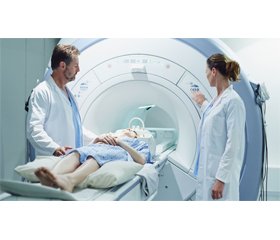
Background. Outlet obstruction syndrome refers to all pelvic floor dysfunctions that are responsible for incomplete evacuation of fecal contents from the rectum. Defecography is the first step for diagnosis of outlet obstruction syndrome. The free selection of imaging planes, good temporal resolution, and excellent soft tissue contrast has helped to transform this method into the preferred imaging modality for evaluation of patients with pelvic floor dysfunction. The purpose of our study was to compare conventional and magnetic resonance (MR) defecography in patients who were admitted for outlet obstruction syndrome. Materials and methods. Twenty-eight patients who presented with constipation between January 2015 and January 2020 were included in this study. MR defecography was performed 1 to 2 weeks after conventional defecography. The methods were compared with regard to their ability to diagnose anterior rectocele, internal mucosal intussusception with or without rectocele, and puborectal spasm. Additional abnormalities were also noted. Results. Comparison of conventional and MR defecography for their ability to diagnose anterior rectocele, internal mucosal intussusception, and puborectal spasm showed no significant differences between the 2 methods. The continuity correction ratio of the 2 methods for diagnosis of anterior rectocele, internal mucosal intussusception, and puborectal spasm was 0.146, 0.007, and 1.000, respectively. Conclusions. Although conventional defecography is the gold standard for diagnosis of rectocele, intussusception, and puborectal spasm, MR defecography has garnered considerable attention due to the lower radiation, increased safety, and higher incidence for diagnosis of another pathology, such as uterine diversion.
магнітно-резонансна дефекографія; звичайна дефекографія; дисфункція тазового дна; ректоцеле; спазм прямої кишки
магнитно-резонансная дефекография; обычная дефекография; дисфункция тазового дна; ректоцеле; спазм прямой кишки
magnetic resonance defecography; conventional defecography; outlet obstructive syndrome; rectocele; puborectal spasm
Introduction
Materials and methods
Results
Discussion
Conclusions
- Jones R., Lydeard S. Irritable bowel syndrome in the general population. Br. Med. J. 1992. 304. Р. 87-90. doi: 10.1136/bmj.304.6819.87.
- Talley N.J., Zinsmeister A.R., Van Dyke C., Melton L.J. 3rd. Epidemiology of colonic symptoms and the irritable bowel syndrome. Gastroenterology. 1991. 101. Р. 927-934. doi: 10.5555/uri:pii:001650859190717Y.
- D’Hoore A., Penninckx F. Obstructed defecation. Colorectal Dis. 2003. 5(4). Р. 280-287. doi: 10.1046/j.1463-1318.2003.00497.x.
- Faccioli N., Comai A., Mainardi P., Perandini S., Moore F., Pozzi-Mucelli R. Defecography: a practical approach. Diagn Interv Radiol. 2010. 16(3). Р. 209-216. doi: 10.4261/1305-3825.DIR.2584-09.1.
- Martín-Martín G.P., García-Armengol J., Roig-Vila J.V., Espí-Macías A., Martínez-Sanjuán V., Mínguez-Pérez M. et al. Magnetic resonance defecography versus videodefecography in the study of obstructed defecation syndrome: Is videodefecography stil the test of choice after 50 years? Tech. Coloproctol. 2017. 21(10). Р. 795-802. doi: 10.1007/s10151-017-1666-0.
- Foti P.V., Farina R., Riva G., Coronella M., Fisichella E., Palmucci S. et al. Pelvic floor imaging: comparison between magnetic resonance imaging and conventional defecography in studying outlet obstruction syndrome. Radiol. Med. 2013. 118. Р. 23-39. doi: 10.1007/s11547-012-0840-8.
- Pannu H.K., Kaufman H.S., Cundiff G.W., Genadry R., Bluemke D.A., Fishman E.K. et al. Dynamic MR imaging of pelvic organ prolapse: spectrum of abnormalities. Radiographics. 2000. 20. Р. 1567-1582. doi: 10.1148/radiographics.20.6.g00nv311567.
- Piloni V., Bergamasco M., Melara G., Garavello P. The clinical value of magnetic resonance defecography in males with obstructed defecation syndrome. Tech. Coloproctol. 2018. 22. Р. 179-190. doi: 10.1007/s10151-018-1759-4.
- Shorvon P.J., McHugh S., Diamant N.E., Somers S., Stevenson G.W. et al. Defecography in normal volunteers: results and implications. Gut. 1989. 30. Р. 1737-1749. doi: 10.1136/gut.30.12.1737.
- Siproudhis L., Dautreme S., Ropert A., Bretagne J.F., Heresbach D., Raoul J.L. et al. Dyschezia and rectocele — a marriage of convenience? Physiologic evaluation of the rectocele in a group of 52 women complaining of difficulty in evacuation. Dis. Colon. Rectum. 1993. 36(11). Р. 1030-1036. doi: 10.1007/BF02047295.
- Pfeifer J., Oliveira L., Park U.C., Gonzalez A., Agachan F., Wexner S.D. et al. Are interpretations of video defecographies reliable and reproducible? Int. J. Colorectal. Dis. 1997. 12. Р. 67-72. doi: 10.1007/s003840050083.
- Johansson C., Nilsson B.Y., Mellgren A., Dolk A., Holmström B. Paradoxical sphincter reaction and associated colorectal disorders. Int. J. Colorectal Dis. 1992. 7. Р. 89-94. doi: 10.1007/BF00341293.
- Bharucha A.E., Wald A., Enck P., Rao S. Functional anorectal disorders. Gastroenterology. 2006. 130. Р. 1510-1518. doi: 10.1053/j.gastro.2005.11.064.
- Halligan S., Malouf A., Bartram C.I., Marshall M., Hollings N., Kamm M.A. Predictive value of impaired evacuation at proctography in diagnosing anismus. Am. J. Roentgenol. 2001. 177. Р. 633-636. doi: 10.2214/ajr.177.3.1770633.
- Roos J.E., Weishaupt D., Wildermuth S., Willmann J.K., Marincek B., Hilfiker P.R. Experience of 4 years with open MR defecography: pictorial review of anorectal anatomy and disease. Radiographics. 2002. 22. Р. 817-832. doi: 10.1148/radiographics.22.4. g02jl02817.
- Reiner C.S., Tutuian R., Solopova A.E., Pohl D., Marincek B., Weishaupt D. MR defecography in patients with dyssynergic defecation: spectrum of imaging findings and diagnostic value. The British Journal of Radiology. 2011. 84. Р. 136-144. doi: 10.1259/bjr/28989463.
- Marzena N., Anna G., Anita K.L., Paweł N. Rectal duplication with sciatic hernia. Videosurgery Miniinv. 2015. 10. Р. 282-285. doi: 10.5114/wiitm.2015.52708.
- Bertschinger K.M., Hetzer F.H., Roos J.E., Treiber K., Marincek B., Hilfiker P.R. Dynamic MR imaging of the pelvic flor performed with patient sitting in an open-magnet unit versus with patient supine in a closed-magnet unit. Radiology. 2002. 223. Р. 501-508. doi: 10.1148/radiol.2232010665.
- Bolog N., Weishaupt D. Dynamic MR Imaging of Outlet Obstruction. Romanian Journal of Gastroenterology. 2005. 14. Р. 293-302. PMID: 16200243.
- Delemarre J.B., Kruyt R.H., Doornbos J., Trimbos J.B., Hermans J., Gooszen H.G. Anterior rectocele: assessment with radiographic defecography, dynamic magnetic resonance imaging, and physical examination. Dis. Colon. Rectum. 1994. 37. Р. 249-259. doi: 10.1007/BF02048163.
- Vanbeckevoort D., Van Hoe L., Oyen R., Ponette E., De Ridder D., Deprest J. Pelvic floor descent in females: comparative study of colpocystodefecography and dynamic fast MR imaging. J. Magn. Reson. Imaging. 1999. 9. Р. 373-377. doi: 10.1002/(SICI)1522-2586(199903)9:3<373::AID-JMRI2>3.0.CO;2-H.
- Vitton V., Vignally P., Barthet M., Cohen V., Durieux O., Bouvier M. et al. Dynamic anal endosonography and MRI defecography in diagnosis of pelvic floor disorders: comparison with conventional defecography. Dis. Colon. Rectum. 2011. 54. Р. 1398-1404. doi: 10.1097/DCR.0b013e31822e89bc.
- Cappabianca S., Reginelli A., Iacobellis F., Granata V., Urciuoli L., Alabiso M.E. et al. Dynamic MRI defecography vs. entero-colpo-cystodefecography in the evaluation of midline pelvic floor hernias in female pelvic floor disorders. Int. J. Colorectal. Dis. 2011. 26. Р. 1191-1196. doi: 10.1007/s00384-011-1218-4.
- Schreyer A.G., Paetzel C., Fürst A., Dendl L.M., Hutzel E., Müller-Wille R. et al. Dynamic magnetic resonance defecography in 10 asymptomatic volunteers. World J. Gastroenterol. 2012. 18. Р. 6836-6842. doi: 10.3748/wjg.v18.i46.6836.


/38.jpg)