Інформація призначена тільки для фахівців сфери охорони здоров'я, осіб,
які мають вищу або середню спеціальну медичну освіту.

Підтвердіть, що Ви є фахівцем у сфері охорони здоров'я.


UkrainePediatricGlobal

UkrainePediatricGlobal

Журнал «Здоровье ребенка» Том 15, №3, 2020

Вернуться к номеру

Синдром ятагана: подходы к диагностике и ведению

Авторы: I.Yu. Avramenko, N.S. Kosmynina
Danylo Halytsky Lviv National Medical University, Lviv, Ukraine

Рубрики: Педиатрия/Неонатология

Разделы: Справочник специалиста

Версия для печати


Резюме

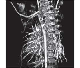
У статті подано клінічний випадок синдрому ятагана в дівчинки віком 3 роки. Захворювання маніфестувало у місячному віці з блідості, перiорального ціанозу та епізоду апное. Дитина була госпіталізована для подальшого обстеження. У дівчинки спостерігався перiоральний ціаноз, SpO2 90 %, частота дихальних рухів 36/хв. Аускультативно в легенях пуерильне дихання, ослаблене справа по передній поверхні грудної клітки. Серцеві тони ритмічні, систолічний шум 2/6 угорі лівого краю грудини і 4/6 у міжлопатковій ділянці зліва. При ехокардіографії діагностовано некритичну коарктацію аорти, дефект міжпередсердної перетинки. При рентгенографії органів грудної клітки в ділянці правого легеневого поля інтенсивна тінь від верхівки до рівня V ребра з нечіткими краями. Тінь серця зміщена вправо. Що в дитини: декстрокардія, декстрапозиція, пневмонія, ателектаз, тимо­мегалія? При бронхоскопії виявлено обструкцію бронхів верхньої та середньої долі правої легені з густим харкотинням білого кольору. Проведено санацію. Після бронхоскопії нормалізувалася сатурація, але, незважаючи на ліквідацію причини ателектазу, контрольна рентгенографія залишалася без змін. Проведено комп’ютерну томографію органів грудної порожнини з контрастуванням. У дитини діагностовано синдром ятагана в поєднанні з коарктацією аорти і аберантною правою підключичною артерією. У віці 2,5 міс. виконано балонну дилатацію коарктації аорти. У віці 2 років проведено хірургічну корекцію — усунення аномального дренажу легеневих вен та пластику дуги аорти. Інтраопераційна анатомія: мезокардія, праві відділи серця дилатовані, нижня доля правої легені гіпоплазована. Пацієнтка фактично не мала симптомів, характерних для синдрому ятагана, оскільки були відсутні ознаки серцевої недостатності, дівчинка не хворіла на респіраторні захворювання. Діагноз у грудному віці було встановлено завдяки епізоду аспірації, але ателектаз і мезокардія завадили візуалізації характерної рентгенологічної ознаки синдрому ятагана. Тому повне дослідження з використанням комп’ютерної томографії з контрастуванням є обов’язковим для таких пацієнтів.

В статье представлен клинический случай синдрома ятагана у 3-летней девочки. Заболевание манифестировало в месячном возрасте с бледности, периорального цианоза и эпизода апноэ. Ребенок госпитализирован для дальнейшего обследования. У девочки наблюдался пери­оральный цианоз, SpO2 90 %, частота дыхательных движений 36/мин. Аускультативно в легких пуэрильное дыхание, ­ослабленное справа по передней поверхности грудной клетки. Сердечные тоны ритмичные, систолический шум 2/6 вверху левого края грудины и 4/6 в межлопаточной области слева. При эхокардиографии диагностированы некритическая коарктация аорты, дефект межпредсердной перегородки. При рентгенографии органов грудной клетки в области правого легочного поля интенсивная тень от верхушки до уровня V ребра с нечеткими краями. Тень сердца смещена вправо. Что у ребенка: декстрокардия, декстрапозиция, пневмония, ателектаз, тимомегалия? При бронхоскопии обнаружена обструкция бронхов верхней и средней доли правого легкого с густой мокротой белого цвета. Проведена санация. После бронхоскопии нормализовалась сатурация, но, несмотря на ликвидацию причины ателектаза, контрольная рентгенография оставалась без изменений. Проведена компьютерная томография органов грудной полости с контрастированием. У ребенка диагностирован синдром ятагана в сочетании с коарктацией аорты и аберрантной правой подключичной артерией. В возрасте 2,5 мес. выполнена баллонная дилатация коарктации аорты. В возрасте 2 лет проведена хирургическая коррекция — устранение аномального дренажа легочных вен и пластика дуги аорты. Интраоперационная анатомия: мезокардия, правые отделы сердца дилатированы, нижняя доля правого легкого гипоплазирована. Наша пациентка фактически не имела симптомов, характерных для синдрома ятагана, поскольку отсутствовали признаки сердечной недостаточности, девочка не болела респираторными заболеваниями. Диагноз в грудном возрасте был установлен благодаря эпизоду аспирации, но ателектаз и мезокардия помешали визуализации характерной рентгенологической картины синдрома ятагана. Поэтому полное исследование с использованием компьютерной томографии с контрастированием является обязательным для таких пациентов.

The article presents a clinical case of scimitar syndrome in a child, who is 3 years old. The manifestation of clinical signs was observed in a child at the age of one month as paleness, perioral cyanosis and episode of apnea. This child was hospitalized for further examination. The girl had perioral cyanosis, SpO2 90 %, respiratory rate is 36/min. Puerile breathing is auscultative in the lungs, and weakened on the right, on the anterior surface of the chest. Heart tones are rhythmic, systolic murmur is 2/6 at the upper left edge of the chest and 4/6 in the interlobular area on the left. Due to echocardiography it was diagnosed non-critical coarctation of the aorta, a secondary defect of the interventricular septum. Chest X-ray: in the area of the right pulmonary field there is an intense shadow from the apex to the level of the 5th rib with fuzzy edges. What does the child have: dextocardia, dextroposition, pneumonia, atelectasis or thymomegaly? Providing the bronchoscopy, it was revealed the obstruction of the bronchi of the upper and middle parts of the right lung with thick murmur of white color. The bronchial tree was rehabilitated. Saturation was normalized after bronchoscopy. But, despite the elimination of the cause of atelectasis, the control radiography was still unchanged. For further diagnosis, CT scan of the chest with contrast was performed. The child was diagnosed with scimitar syndrome in combination with aortic coarctation and aberrant right subclavian artery. Balloon dilation of aortic coagulation was performed at the age of 2.5 months. At the age of 2 years, surgical correction was performed — elimination of abnormal drainage of the pulmonary veins and the aortic arch plastic. Intraoperative anatomy: mesocardia, right heart sections are dilated, lower part of the right lung is hypoplasic. Our patient did not actually have symptoms characteristic of the scimitar syndrome because there were no signs of heart failure, the girl did not have respiratory disease. A diagnosis for a baby at infancy was made due to an aspiration episode. But atelectasis and mesocardia prevented visualization of the characteristic radiological feature of scimitar syndrome. Therefore, a complete study using contrast-enhanced CT is mandatory for such patients.


Ключевые слова

синдром ятагана; діти; діагностика

синдром ятагана; дети; диагностика

scimitar syndrome; children; diagnosis

Despite the continuous improvement of the methods of prenatal diagnosis of fetal malformations, the limited possibilities of intrauterine verification of vascular anomalies of the lungs still take place. One of the most rare and difficult to diagnose forms of combination of vascular anomalies is partial abnormal pulmonary vein drainage, which is sometimes a component of scimitar syndrome (ICD-10: Q26.8) [1].
Scimitar syndrome (hypogenetic pulmonary syndrome, venolobar syndrome or “yataghan” syndrome) belongs to a group of venolobar syndromes associated with lung sequestration and congenital heart disease. This is a congenital anomaly characterized by the fact that one or more pulmonary veins drain not into the left atrium, as normal, but directly into the inferior vena cava above or below the diaphragm. Scimitar syndrome accounts 4 % of abnormal pulmonary vein drainage. This syndrome is associated with changes in the arterial supply of the lungs, the right lung is hypoplasic, as well as the hypoplasic right bronchus and the right pulmonary artery. As far as the heart is concerned, in addition to dextroposition of the heart with displacement of the mediastinum, the most frequent changes (1/4 cases) are the absence of the pericardium, and other defects of development, such as defect of the atrial (60–70 %) or interventricular septum, coarctation of the aorta and tetralogy of Fallot (25 %).
Scimitar syndrome is a rare congenital pathology and occurs from 1 to 3 cases in 100,000 newborns, predominantly in girls. In 1836 the pathology was first described on an autopsy material by Dr. G. Cooper, who gave it a name because of the chest X-ray: a long and curved shadow on the right contour of the heart is similar to the blade of a yataghan. The morbidity and mortality of this pathology is considered to be low since most cases are identified by accident or at the autopsy [2, 3].
Clinical features with scimitar syndrome vary greatly depending on age. Patients are diagnosed with early (infantile) type or late (infant/adult) type. In the neonatal period, scimitar syndrome is characterized by heart or respiratory failure, secondary pulmonary hypertension, and usually requires surgical treatment and a worse prognosis. There is tachypnea, decreased appetite, such children are lagging behind in physical development. Severe cases lead to cyanosis and pulmonary hypertension, which occurs as a result of constant discharge of blood from left to right due to the abnormal circulation in the right lung, and this leads to the increased pressure in the right parts of the heart. Pulmonary hypoplasia further shortens the circulatory system and, in combination with pulmonary vein obstruction or other congenital malformations, produces persistent pulmonary hypertension. When examined, the heart is shifted to the right, the auscultatory picture of the lungs is not changed or there is weakened breathing. The prognosis depends on the severity of concomitant anomalies, the level of pulmonary hypertension and the child’s clinical condition [4].
Symptoms in the infant/adult type have a milder course. In adults, the defects of short pulmonary circumference are negligible: with no pulmonary hypertension in 77 % of cases or with pulmonary hypertension in 23 %. About a half of the patients diagnosed after their first year of living remain asymptomatic. Partial abnormal drainage of the pulmonary veins creates a left-right shunt, which is often not clinically manifested before the adulthood or is detected accidentally on chest radiographs after recurrent pulmonary infections. This type may be accompanied by fatigue, shortness of breath and recurrent pneumonia. If this defect has not been diagnosed and corrected in time, with age in the patients there are developed volume overload of the right heart departments, pulmonary hypertension, right ventricular failure and tricuspid regurgitation. Surgical treatment in the adult type is recommended to delay as much as possible providing conservative therapy for pulmonary hypertension.
Scimitar syndrome is diagnosed in 70 % of cases by means of echocardiography, which has high sensitivity and allows detecting other cardiac abnormalities. Three-dimensional CT or MRI of the heart is fundamental to visualize abnormal pulmonary vein drainage and other anatomical features of a patient. Cardiac catheterization and angiography confirm the diagnosis, providing detailed information on the anatomy of defects and the severity of pulmonary hypertension that is important for planning surgical correction.
Treatment depends on the patient’s age and symptoms. Newborns are treated for pulmonary hypertension. Surgical treatment is recommended for children with severe heart failure, recurrent respiratory tract infections, or pulmonary hypertension resistant to the therapy. In the case of lung sequestration, lobectomy and blocking of an abnormal systemic flow by embolization or ligation of the server artery are required. Other congenital heart defects (septal defects, aortic coarctation, tetralogy of Fallot) should be clearly corrected. However, most researchers believe that all patients diagnosed with scimitar syndrome need surgical correction to prevent complications with age [5, 6].
We bring to your attention a clinical case of a child with scimitar syndrome.
Olenka D. was born at the second pregnancy (gestation period of 38 weeks), the second physiological birth, birth weight of 3350. And the pregnancy in the mother ended with an early spontaneous miscarriage. She was first referred to a cardiologist at the age of one month because of paleness and perioral cyanosis. Due to echocardiography it was diagnosed non-critical coarctation of the aorta, a secondary defect of the interventricular septum of 0.6 cm and the child was admitted to the hospital for further examination.
The anamnesis shows that apnea and cyanosis were observed 4 days before hospitalization. The baby’s aunt has a two-part right lung.
The child’s condition is considered to be moderate and stable. Weight is 4 kg. Perioral cyanosis, SpO2 90 %, respiratory rate is 36/min. Puerile breathing is auscultative in the lungs, and weakened on the right, on the anterior surface of the chest. Heart tones are rhythmic, systolic murmur is 2/6 at the upper left edge of the chest and 4/6 in the interlobular area on the left. Heart rate is 135/min, blood pressure is: right arm — 83/36 mm Hg, left arm — 80/35 mm Hg and leg — 85/47 mm Hg. Pulsation is weakened on the peripheral arteries. The liver is palpated along the edge of the costal arch. There is no edema.
Results of laboratory tests are within normal limits.
ECG. Sinus rhythm, 135/min, right chart, incomplete blockage of the right leg of the bundle of His.
Echocardiography. Left ventricular: ultimate diastolic size of 1.9 cm. Right ventricular: ultimate diastolic size of 2.3 cm. Left atrium of 0.7 cm. Right atrium of 1.4 cm. Secondary atrial septal defect is 0.6 cm. Atrioventricular valves are unchanged. Ascending aorta of 0.7 cm. Aorta coarctation of 0.3 cm with a pressure gradient of 25 mm Hg. Aberrant right subclavian artery. Pulmonary artery of 1.3 cm, branches of 0.6 cm. Inferior vena cava of 1 cm width with a constant atypical flow. It is not possible to visualize the infusion of the right pulmonary veins into the left atrium.
Chest X-ray. In the area of the right pulmonary field there is an intense shadow from the apex to the level of the 5th rib with fuzzy edges. The shadow of the heart is shifted to the right. On the left contour in the upper mediastinum there is a shadow of the thymus (Fig. 1). Taking into consideration such a radiographic picture, the question is, what does the child have: dextrocardia, dextroposition, pneumonia, atelectasis or thymomegaly? The absence of intoxication and inflammatory changes at the laboratory examination caused a doubt that it is the diagnosis of pneumonia. Taking into account the possible episode of aspiration, the radiographic picture was considered as atelectasis of the right lung with the shift of the mediastinum to the right. It was made a decision to provide a remedial diagnostic bronchoscopy.
Providing the bronchoscopy, it was revealed the obstruction of the bronchi of the upper and middle parts of the right lung with thick murmur of white color. The bronchial tree was rehabilitated. Saturation was normalized after bronchoscopy. But, despite the elimination of the cause of atelectasis, the control radiography was still unchanged.
For further diagnosis, CT scan of the chest with contrast was performed (Fig. 2, 3). Aortic coarctation and aberrant right subclavian artery were confirmed. Right pulmonary veins create a collector of up to 6 mm in size, which drains into the supradiaphragmatic part of the inferior vena cava. Narrowing is visualized in the place of inflow of the collector into the inferior vena cava.
The child was diagnosed with scimitar syndrome in combination with aortic coarctation and aberrant right subclavian artery. Balloon dilation of aortic coarctation was performed at the age of 2.5 months. The girl has been observed by a cardiologist for 2 years. As physical and psychomotor development is concerned, she kept up with her peers. Three times a year she was ill with respiratory di–seases. During the observation, the pressure gradient at the coarctation of the aorta was kept within 10–13 mm Hg. At the age of 2 years, surgical correction was performed — elimination of abnormal drainage of the pulmonary veins and the aortic arch plastic. Intraoperative anatomy: mesocardia, right heart sections are dilated, lower part of the right lung is hypoplasic.
The anatomical data of the child is a variant of scimitar syndrome: infracardiac form of partial abnormal drainage of the pulmonary veins with obstruction of the collector at the inflow into the inferior vena cava in combination with coarctation of the aorta, atrial septal defect, abnormal outflow of the right subclavian artery, and hypoplasia of the lower part of the right lung. The clinical picture of patients with scimitar syndrome is extremely diverse. Recurrent respiratory infections are characteristic of scimitar syndrome. Recurrent pneumonia with scimitar syndrome may result from external airway compression by the collector of the pulmonary veins. Other mechanisms suggest impaired lymphatic drainage and insufficient local activity of lymphocytes and macrophages in the affected lung area [7]. Our patient did not actually have symptoms characteristic of scimitar syndrome because there were no signs of heart failure, the girl did not have respiratory disease. A diagnosis for a baby at infancy was made due to an aspiration episode. But atelectasis and mesocardia prevented visualization of the characteristic radiological feature of scimitar syndrome. Therefore, a complete study using contrast-enhanced CT is mandatory for such patients.
Surgical correction of abnormal pulmonary vein drainage is required for symptoms of heart failure [8, 9]. Instead, children in serious condition may need lobectomy or pneumonectomy [10]. However, it is the most difficult to make decision on treatment (surgical or medical) for children with mild scimitar syndrome, at the asymptomatic course and having a random diagnosis, as in our case described here. For this purpose, it is necessary to observe the development of pulmonary hypertension.
Conflicts of interests. Authors declare the absence of any conflicts of interests and their own financial interest that might be construed to influence the results or interpretation of their manuscript.

Список литературы

  1. Golubeva M.V., Ilyina N.A., Kagan A.V. Features of pulmonary circulation in children with congenital pulmonary venolobar syndrome. Regional blood circulation and microcirculation. 2019. 18(1). 55-65. https://doi.org/10.24884/1682-6655-2019-18-1-55-65.
  2. Icek S., Arslan H., Ugurlucan M., Yildiz Y. Scimitar Syndrome: The Curved Turkish Sabre. Seminars in Thoracic and Cardiovascular Surgery: Pediatric Cardiac Surgery Annual. 2014. 7(1). 56-61. doi: 10.1053/j.pcsu.2014.01.003.
  3. Gustainyte V., Miller M., Towbin T., Towbin A., Neville Kucera J. Scimitar syndrome. Appl. Radiol. 2019. 48(5). 37-39.
  4. Shamrani A., Sadi R., Ihoury M., Harbi A. Infantile scimitar syndrome with unusual associations. Saudi Medical Journal. 2017. 38(7). 764-767. doi: 10.15537/smj.2017.7.18365. PMID: 28674724. PMCID: PMC5556286.
  5. Lark M., Cai A., Rideout P., Gregg D., Suranyi P. Novel Technique of Surgical Management of Scimitar Syndrome. Case Reports in Cardiology. 2019. 5. https://doi.org/10.1155/2019/6932680.
  6. Di Filippo S. Epidemiology and Physiopathology of Scimitar Syndrome. In: The Complete Reference for Scimitar Syndrome. Anatomy, Epidemiology, Diagnosis and Treatment. 2017. 6. 57-66. doi: 10.1016/B978-0-12-810406-4.00005-9.
  7. Jackson N., Nokes B.T., Sakata K., Cummings K., Vaszar L. An unusual variant of scimitar syndrome predisposing to recurrent pneumonia. Respiratory Medicine Case Reports. 2019. 23(26). 240-243. https://doi.org/10.1016/j.rmcr.2019.01.010.
  8. Lugones I., Garcia R. A new surgical approach to scimitar syndrome. Ann. Thorac. Surg. 2014. 97. 353-355. doi: 10.1016/j.athoracsur.2013.05.105.
  9. Brink J., Yong M.S., D’Udekem Y., Weintraub R.G., Brizard C.P., Konstantinov I.E. Surgery for scimitar syndrome: the Melbourne experience. Interact. Cardiovasc. Thorac. Surg. 2015. 20. 31-34. doi: 10.1093/icvts/ivu319. Epub 2014 Oct 6.
  10. Vida V.L., Padalino M.A., Boccuzzo G., Tarja E., Berggren H., Carrel T., Cicek S. et al. Scimitar syndrome: a European Congenital Heart Surgeons Association (ECHSA) multicentric study. Circulation. 2010. 122. 1159-1166. doi: 10.1161/CIRCULATIONAHA.109.926204. Epub 2010 Sep 7.

Вернуться к номеру