Журнал «Травма» Том 17, №6, 2016
Вернуться к номеру
Вивчення напружено-деформованого стану системи «інтрамедулярний фіксатор — уламки» на різних етапах відновлення функції сегмента кінцівки після хірургічних втручань
Авторы: Пашенко А.В.(1, 2), Хмизов С.О.(1), Тяжелов О.А.(1), Карпінський М.Ю.(1), Карпінська О.Д.(1), Яресько О.Д.(1)
(1) — ДУ «Інститут патології хребта та суглобів ім. проф. М.І. Ситенка НАМН України», м. Харків, Україна
(2) — Харківська медична академія післядипломної освіти, м. Харків, Україна
Рубрики: Травматология и ортопедия
Разделы: Клинические исследования
Версия для печати
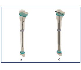
У статті проведено аналіз результатів визначення напружено-деформованого стану біомеханічної системи «інтрамедулярний фіксатор — уламки» на різних етапах відновлення функції сегмента кінцівки після хірургічних втручань. Дослідження проводилося шляхом математичного моделювання методом кінцевих елементів напружено-деформованого стану великогомілкової кістки в умовах недосконалого остеогенезу з використанням інтрамедулярних фіксаторів різних типів — титанових еластичних стрижнів, інтрамедулярного телескопічного фіксатора без ротаційної стабільності, а також ротаційно-стабільного інтрамедулярного телескопічного фіксатора після моделювання коригуючої остеотомії.
В статье проведен анализ результатов определения напряженно-деформированного состояния биомеханической системы «интрамедуллярный фиксатор — отломки» на разных этапах восстановления функции сегмента конечности после хирургических вмешательств. Исследование проводилось путем математического моделирования методом конечных элементов напряженно-деформированного состояния большеберцовой кости в условиях несовершенного остеогенеза с использованием интрамедуллярных фиксаторов различных типов — титановых эластичных стержней, интрамедуллярного телескопического фиксатора без ротационной стабильности, а также ротационно-стабильного интрамедуллярного телескопического фиксатора после моделирования корригирующей остеотомии.
Background. Surgical correction of multiplanar deformities of long bones of the limbs in growing children with osteogenesis imperfecta is one of the most important stages in the treatment of these patients. At the present stage of development of orthopedic surgery, intramedullary telescopic nails of different types are being used in growing children with osteogenesis imperfecta. Objective: to determine the stress-strain state of the system «intramedullary fixator — fragments» in various stages of the recovery of limb segment function after surgery. Materials and methods. In modeling the union, mechanical properties of this element were replaced by the properties of cortical bone. In the simulation of «bone — implant» system, this item increased by 5 cm during the growth. Material was considered homogeneous and isotropic. As the final element, 10-node tetrahedron with quadratic approximation has been chosen. Mechanical properties of healthy bone were chosen according to V.P. Agapov, V.A. Berezovsky, mechanical properties of the bone tissue in oseogenesis imperfecta — according to Z.F. Fan and J.M. Fritz, and the characteristics of synthetic materials — according to J.M. Gere, S.P. Timoshenko. We used characteristics, such as E — elastic modulus (Young’s modulus), ν — Poisson’s ratio. The value of the load on the compression and flexion was 350 N, corresponding to the load of the body of a child weighing 50 kg (500 N) at one-leg standing (without the weight of standing leg). When studying models for torsion, torque moment of 10 Nm is applied to the proximal articular surface. The magnitude of the stress has been controlled in 11 areas: 1–10 — checkpoints, 11 — zone of maximum stress on the metal structure. Results. The first phase of the study: elastic titanium rods come under very considerable strain for all types of stress that can lead to their destruction or deformation, but it does not lead to the discharge of bone tissue, which is a negative factor in terms of impaired bone quality. Intramedullary telescopic nails with rotational stability works well under the influence of torsion loads that can significantly unload bone tissue, especially around areas of growth. The second phase of the study: forming a complete bone regenerate leading to inclusion in the process of loading, which leads to even distribution of stresses in the bone for all types of stress, and unloading the fixation devices. With rotary load, rotary stable intramedullary telescopic nail continues to serve as internal «splinting» of the bone. The third phase of the study: the increased length of the bone after completion of its growth does not lead to significant changes in the distribution of stresses in the models. Conclusions. Intramedullary telescopic nails with rotational stability under load torsion work more effectively for all other types of implants. Their use can reduce and evenly distribute stress over the entire length of the bone.
напружено-деформований стан кістки; інтрамедулярний телескопічний фіксатор
напряженно-деформированное состояние кости; интрамедуллярный телескопический фиксатор
stress-strain state of the bone; intramedullary telescopic nail
Статтю опубліковано на с. 62-75
Вступ
Матеріали та методи
/63.jpg)
Результати
Стабілізація після зрощення
Після завершення росту
Висновки
1. Bailey R.W., Dubow H.I. Studies of longitudinal bone growth resulting in an extensible nail // Surg. Forum. — 1963. — 14. — 455-458;
2. Bailey R.W., Dubow H.I. Evolution of the concept of an extensible nail accommodating to normal longitudinal bone growth: clinical considerations and implications // Clin. Orthop. — 1981. — 159. — 157-169.
3. Elena Monti, Monica Mottes, Paolo Fraschini et al. Current and emerging treatments for the management of osteogene-sis imperfect // Therapeutics and Clinical Risk Management. — 2010. — 6. — 367-381.
4. Francois Fassier, Pierre Duval, Ariel Dujovne. Intamedullary nail system. — Patent № US 06524213. — Feb. 25 2003.
5. KAN Saldanha, Saleh М., Bell M.J., Fernandes J.A. Limb Reconstruction on Osteogenesis Imperfecta // J. Bone Joint Surg. Br. — 2003. — Vol. 85-B.
6. Navin N. Thakkar. Flexible nail assembly for fracturesof long bones. — Patent № US 2007/0173834. — Jul. 26 2007.
7. Агапов В.П. Метод конечных элементов в статике, динамике и устойчивости пространственных тонкостенных подкрепленных конструкций: Уч. пособие / В.П. Агапов. — М.: Изд. АСВ, 2000. — 152 с.
8. Березовский В.А., Колотилов Н.Н. Биофизические характеристики тканей человека: Справочник. — К.: Наукова думка, 1990. — 224 с.
9. Fan Z.F. et al. Connective Tissue Research. — 2007. — 48. — 70-75.
10. Fritz J.M. et al. Medical Engineering & Physics. — 2009. — Vol. 31. — Р. 1043-1048.
11. Gere J.M., Timoshenko S.P. Mechanics of Material. — 1997. — 912 c.
12. Кнетс И.В., Пфафрод Г.О., Саулгозис Ю.Ж. Деформирование и разрушение твердых биологических тканей. — Рига: Зинатне, 1980. — 320с.
13. Образцов И.Ф., Адамович И.С., Барер И.С. и др. Проблема прочности в биомеханике: Учебное пособие для технич. и биол. спец. вузов. — М.: Высш. школа, 1988. — 311 с.
14. Зенкевич О.К. Метод конечных элементов в технике. — М: Мир, 1978. — 519 с.
15. Алямовский А.А. SolidWorks/COSMOSWorks. Инженерный анализ методом конечных элементов / А.А. Алямовский. — М.: ДМК Пресс, 2004. — 432 с.


/63_2.jpg)
/63_3.jpg)
/64.jpg)
/64_2.jpg)
/65.jpg)
/68_2.jpg)
/69.jpg)
/70.jpg)
/66.jpg)
/71.jpg)
/72.jpg)
/73.jpg)
/67.jpg)
/68.jpg)